1ZL7

Crystal structure of catalytically-active phospholipase A2 with bound calcium


Experimental Data Snapshot

  • Method: X-RAY DIFFRACTION
  • Resolution: 1.60 Å
  • R-Value Free: 0.232 
  • R-Value Work: 0.176 
  • R-Value Observed: 0.179 

wwPDB Validation   3D Report Full Report


This is version 1.4 of the entry. See complete history


Literature

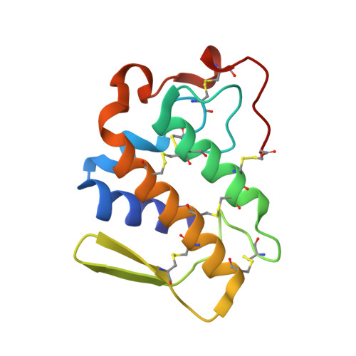
Insights into metal ion binding in phospholipases A(2): ultra high-resolution crystal structures of an acidic phospholipase A(2) in the Ca(2+) free and bound states.

Murakami, M.T.Gabdoulkhakov, A.Genov, N.Cintra, A.C.Betzel, C.Arni, R.K.

(2006) Biochimie 88: 543-549

  • DOI: https://doi.org/10.1016/j.biochi.2005.10.014
  • Primary Citation of Related Structures:  
    1ZL7, 1ZLB

  • PubMed Abstract: 

    The electrophile Ca(2+) is an essential multifunctional co-factor in the phospholipase A(2) mediated hydrolysis of phospholipids. Crystal structures of an acidic phospholipase A(2) from the venom of Bothrops jararacussu have been determined both in the Ca(2+) free and bound states at 0.97 and 1.60 A resolutions, respectively. In the Ca(2+) bound state, the Ca(2+) ion is penta-coordinated by a distorted pyramidal cage of oxygen and nitrogen atoms that is significantly different to that observed in structures of other Group I/II phospholipases A(2). In the absence of Ca(2+), a water molecule occupies the position of the Ca(2+) ion and the side chain of Asp49 and the calcium-binding loop adopts a different conformation.


  • Organizational Affiliation

    Department of Physics, IBILCE/UNESP, Cristovão Colombo 2265, São José do Rio Preto, SP 15054-000, Brazil.


Macromolecules
Find similar proteins by:  (by identity cutoff)  |  3D Structure
Entity ID: 1
MoleculeChains Sequence LengthOrganismDetailsImage
hypotensive phospholipase A2122Bothrops jararacussuMutation(s): 0 
EC: 3.1.1.4
UniProt
Find proteins for Q8AXY1 (Bothrops jararacussu)
Explore Q8AXY1 
Go to UniProtKB:  Q8AXY1
Entity Groups  
Sequence Clusters30% Identity50% Identity70% Identity90% Identity95% Identity100% Identity
UniProt GroupQ8AXY1
Sequence Annotations
Expand
  • Reference Sequence
Experimental Data & Validation

Experimental Data

  • Method: X-RAY DIFFRACTION
  • Resolution: 1.60 Å
  • R-Value Free: 0.232 
  • R-Value Work: 0.176 
  • R-Value Observed: 0.179 
  • Space Group: C 2 2 21
Unit Cell:
Length ( Å )Angle ( ˚ )
a = 39.206α = 90
b = 53.218β = 90
c = 90.057γ = 90
Software Package:
Software NamePurpose
REFMACrefinement
MAR345data collection
SCALEPACKdata scaling
AMoREphasing

Structure Validation

View Full Validation Report


Entry History 

Deposition Data

Revision History  (Full details and data files)

  • Version 1.0: 2006-04-25
    Type: Initial release
  • Version 1.1: 2008-04-30
    Changes: Version format compliance
  • Version 1.2: 2011-07-13
    Changes: Derived calculations, Version format compliance
  • Version 1.3: 2017-10-11
    Changes: Refinement description
  • Version 1.4: 2023-08-23
    Changes: Data collection, Database references, Derived calculations, Refinement description