The three-dimensional solution structure of Ca(2+)-bound S100A1 as determined by NMR spectroscopy
Wright, N.T., Varney, K.M., Ellis, K.C., Markowitz, J., Gitti, R.K., Zimmer, D.B., Weber, D.J.(2006) J Mol Biol 353: 410-426
- PubMed: 16169012
- DOI: https://doi.org/10.1016/j.jmb.2005.08.027
- Primary Citation of Related Structures:
1ZFS - PubMed Abstract:
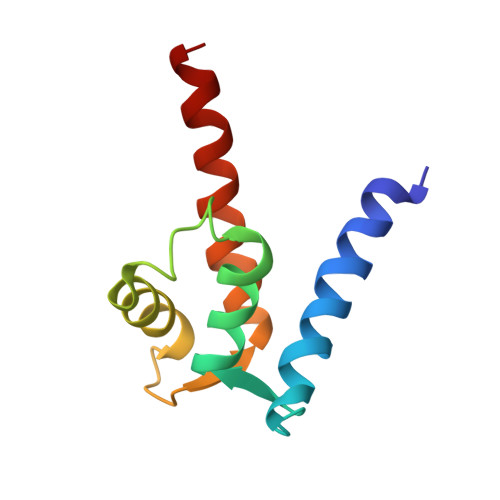
S100A1 is an EF-hand-containing Ca(2+)-binding protein that undergoes a conformational change upon binding calcium as is necessary to interact with protein targets and initiate a biological response. To better understand how calcium influences the structure and function of S100A1, the three-dimensional structure of calcium-bound S100A1 was determined by multidimensional NMR spectroscopy and compared to the previously determined structure of apo. In total, 3354 nuclear Overhauser effect-derived distance constraints, 240 dihedral constraints, 160 hydrogen bond constraints, and 362 residual dipolar coupling restraints derived from a series of two-dimensional, three-dimensional, and four-dimensional NMR experiments were used in its structure determination (>21 constraints per residue). As with other dimeric S100 proteins, S100A1 is a symmetric homodimer with helices 1, 1', 4, and 4' associating into an X-type four-helix bundle at the dimer interface. Within each subunit there are four alpha-helices and a short antiparallel beta-sheet typical of two helix-loop-helix EF-hand calcium-binding domains. The addition of calcium did not change the interhelical angle of helices 1 and 2 in the pseudo EF-hand significantly; however, there was a large reorientation of helix 3 in the typical EF-hand. The large conformational change exposes a hydrophobic cleft, defined by residues in the hinge region, the C terminus, and regions of helix 3, which are important for the interaction between S100A1 and a peptide (TRTK-12) derived from the actin-capping protein CapZ.
Organizational Affiliation:
Department of Biochemistry and Molecular Biology, University of Maryland School of Medicine, 108 N. Greene St., Baltimore, MD 21201, USA.