1YWH

crystal structure of urokinase plasminogen activator receptor


Experimental Data Snapshot

  • Method: X-RAY DIFFRACTION
  • Resolution: 2.70 Å
  • R-Value Free: 0.315 
  • R-Value Work: 0.245 
  • R-Value Observed: 0.249 

wwPDB Validation   3D Report Full Report


Ligand Structure Quality Assessment 


This is version 2.0 of the entry. See complete history


Literature

Crystal structure of the human urokinase plasminogen activator receptor bound to an antagonist peptide

Llinas, P.Le Du, M.H.Gardsvoll, H.Dano, K.Ploug, M.Gilquin, B.Stura, E.A.Menez, A.

(2005) EMBO J 24: 1655-1663

  • DOI: https://doi.org/10.1038/sj.emboj.7600635
  • Primary Citation of Related Structures:  
    1YWH

  • PubMed Abstract: 

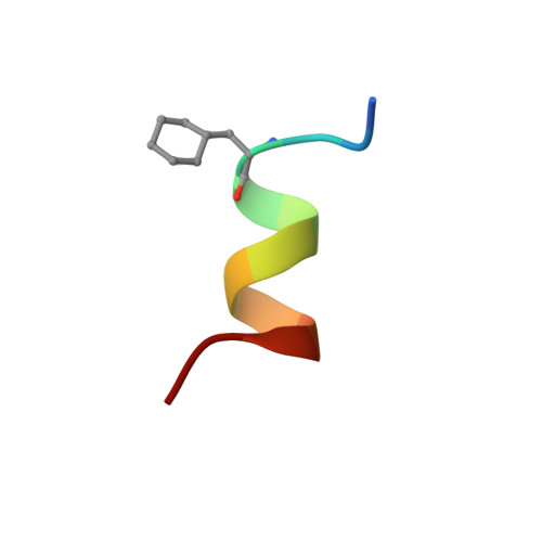
    We report the crystal structure of a soluble form of human urokinase-type plasminogen activator receptor (uPAR/CD87), which is expressed at the invasive areas of the tumor-stromal microenvironment in many human cancers. The structure was solved at 2.7 A in association with a competitive peptide inhibitor of the urokinase-type plasminogen activator (uPA)-uPAR interaction. uPAR is composed of three consecutive three-finger domains organized in an almost circular manner, which generates both a deep internal cavity where the peptide binds in a helical conformation, and a large external surface. This knowledge combined with the discovery of a convergent binding motif shared by the antagonist peptide and uPA allowed us to build a model of the human uPA-uPAR complex. This model reveals that the receptor-binding module of uPA engages the uPAR central cavity, thus leaving the external receptor surface accessible for other protein interactions (vitronectin and integrins). By this unique structural assembly, uPAR can orchestrate the fine interplay with the partners that are required to guide uPA-focalized proteolysis on the cell surface and control cell adhesion and migration.


  • Organizational Affiliation

    CEA, Département d'Ingénierie et d'Etudes des Protéines, CE Saclay, Gif sur Yvette, France.


Macromolecules
Find similar proteins by:  (by identity cutoff)  |  3D Structure
Entity ID: 1
MoleculeChains Sequence LengthOrganismDetailsImage
Urokinase plasminogen activator surface receptor
A, C, E, G, I
A, C, E, G, I, K, M, O
313Homo sapiensMutation(s): 0 
Gene Names: PLAURMO3UPAR
UniProt & NIH Common Fund Data Resources
Find proteins for Q03405 (Homo sapiens)
Explore Q03405 
Go to UniProtKB:  Q03405
PHAROS:  Q03405
GTEx:  ENSG00000011422 
Entity Groups  
Sequence Clusters30% Identity50% Identity70% Identity90% Identity95% Identity100% Identity
UniProt GroupQ03405
Sequence Annotations
Expand
  • Reference Sequence

Find similar proteins by:  Sequence   |   3D Structure  

Entity ID: 2
MoleculeChains Sequence LengthOrganismDetailsImage
antagonist peptide
B, D, F, H, J
B, D, F, H, J, L, N, P
13N/AMutation(s): 0 
Entity Groups  
Sequence Clusters30% Identity50% Identity70% Identity90% Identity95% Identity100% Identity
Sequence Annotations
Expand
  • Reference Sequence
Oligosaccharides

Help

Entity ID: 3
MoleculeChains Length2D Diagram Glycosylation3D Interactions
2-acetamido-2-deoxy-beta-D-glucopyranose-(1-4)-[alpha-L-fucopyranose-(1-6)]2-acetamido-2-deoxy-beta-D-glucopyranoseBA [auth b],
Q,
S,
T
3N-Glycosylation
Glycosylation Resources
GlyTouCan:  G21290RB
GlyCosmos:  G21290RB
GlyGen:  G21290RB
Entity ID: 4
MoleculeChains Length2D Diagram Glycosylation3D Interactions
alpha-L-fucopyranose-(1-6)-2-acetamido-2-deoxy-beta-D-glucopyranose
AA [auth a],
DA [auth d],
GA [auth g],
R,
U,
AA [auth a],
DA [auth d],
GA [auth g],
R,
U,
V,
X,
Y
2N-Glycosylation
Glycosylation Resources
GlyTouCan:  G86851RC
GlyCosmos:  G86851RC
GlyGen:  G86851RC
Entity ID: 5
MoleculeChains Length2D Diagram Glycosylation3D Interactions
2-acetamido-2-deoxy-beta-D-glucopyranose-(1-4)-2-acetamido-2-deoxy-beta-D-glucopyranoseFA [auth f],
W
2N-Glycosylation
Glycosylation Resources
GlyTouCan:  G42666HT
GlyCosmos:  G42666HT
GlyGen:  G42666HT
Entity ID: 6
MoleculeChains Length2D Diagram Glycosylation3D Interactions
alpha-D-mannopyranose-(1-3)-[alpha-D-mannopyranose-(1-6)]beta-D-mannopyranose-(1-4)-2-acetamido-2-deoxy-beta-D-glucopyranose-(1-4)-2-acetamido-2-deoxy-beta-D-glucopyranoseCA [auth c],
EA [auth e],
HA [auth h],
Z
5N-Glycosylation
Glycosylation Resources
GlyTouCan:  G22768VO
GlyCosmos:  G22768VO
GlyGen:  G22768VO
Small Molecules
Ligands 2 Unique
IDChains Name / Formula / InChI Key2D Diagram3D Interactions
NAG
Query on NAG

Download Ideal Coordinates CCD File 
BB [auth I]
FB [auth K]
GB [auth K]
IA [auth A]
JA [auth A]
BB [auth I],
FB [auth K],
GB [auth K],
IA [auth A],
JA [auth A],
JB [auth M],
MB [auth O],
NA [auth C],
OA [auth C],
SA [auth E],
WA [auth G]
2-acetamido-2-deoxy-beta-D-glucopyranose
C8 H15 N O6
OVRNDRQMDRJTHS-FMDGEEDCSA-N
SO4
Query on SO4

Download Ideal Coordinates CCD File 
AB [auth G]
CB [auth I]
DB [auth I]
EB [auth I]
HB [auth K]
AB [auth G],
CB [auth I],
DB [auth I],
EB [auth I],
HB [auth K],
IB [auth K],
KA [auth A],
KB [auth M],
LA [auth A],
LB [auth M],
MA [auth A],
NB [auth O],
PA [auth C],
QA [auth C],
RA [auth C],
TA [auth E],
UA [auth E],
VA [auth E],
XA [auth G],
YA [auth G],
ZA [auth G]
SULFATE ION
O4 S
QAOWNCQODCNURD-UHFFFAOYSA-L
Modified Residues  1 Unique
IDChains TypeFormula2D DiagramParent
ALC
Query on ALC
B, D, F, H, J
B, D, F, H, J, L, N, P
L-PEPTIDE LINKINGC9 H17 N O2ALA
Experimental Data & Validation

Experimental Data

  • Method: X-RAY DIFFRACTION
  • Resolution: 2.70 Å
  • R-Value Free: 0.315 
  • R-Value Work: 0.245 
  • R-Value Observed: 0.249 
  • Space Group: P 1 21 1
Unit Cell:
Length ( Å )Angle ( ˚ )
a = 106.928α = 90
b = 136.831β = 97.27
c = 140.536γ = 90
Software Package:
Software NamePurpose
REFMACrefinement
HKL-2000data reduction
SCALEPACKdata scaling
MOLREPphasing

Structure Validation

View Full Validation Report


Ligand Structure Quality Assessment 


Entry History 

Deposition Data

Revision History  (Full details and data files)

  • Version 1.0: 2005-05-10
    Type: Initial release
  • Version 1.1: 2007-10-16
    Changes: Version format compliance
  • Version 1.2: 2011-07-13
    Changes: Derived calculations, Version format compliance
  • Version 1.3: 2018-01-31
    Changes: Experimental preparation
  • Version 2.0: 2020-07-29
    Type: Remediation
    Reason: Carbohydrate remediation
    Changes: Advisory, Atomic model, Data collection, Database references, Derived calculations, Non-polymer description, Structure summary