Structural and biochemical implications of single amino acid substitutions in the nucleotide-dependent switch regions of the nitrogenase Fe protein from Azotobacter vinelandii
Jang, S.B., Jeong, M.S., Seefeldt, L.C., Peters, J.W.(2004) J Biol Inorg Chem 9: 1028-1033
- PubMed: 15549494
- DOI: https://doi.org/10.1007/s00775-004-0605-5
- Primary Citation of Related Structures:
1XD8, 1XD9, 1XDB - PubMed Abstract:
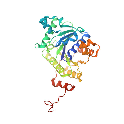
The structures of nitrogenase Fe proteins with defined amino acid substitutions in the previously implicated nucleotide-dependent signal transduction pathways termed switch I and switch II have been determined by X-ray diffraction methods. In the Fe protein of nitrogenase the nucleotide-dependent switch regions are responsible for communication between the sites responsible for nucleotide binding and hydrolysis and the [4Fe-4S] cluster of the Fe protein and the docking interface that interacts with the MoFe protein upon macromolecular complex formation. In this study the structural characterization of the Azotobacter vinelandii nitrogenase Fe protein with Asp at position 39 substituted by Asn in MgADP-bound and nucleotide-free states provides an explanation for the experimental observation that the altered Fe proteins form a trapped complex subsequent to a single electron transfer event. The structures reveal that the substitution allows the formation of a hydrogen bond between the switch I Asn39 and the switch II Asp125. In the structure of the native enzyme the analogous interaction between the side chains of Asp39 and Asp125 is precluded due to electrostatic repulsion. These results suggest that the electrostatic repulsion between Asp39 and Asp125 is important for dissociation of the Fe protein:MoFe protein complex during catalysis. In a separate study, the structural characterization of the Fe protein with Asp129 substituted by Glu provides the structural basis for the observation that the Glu129-substituted variant in the absence of bound nucleotides has biochemical properties in common with the native Fe protein with bound MgADP. Interactions of the longer Glu side chain with the phosphate binding loop (P-loop) results in a similar conformation of the switch II region as the conformation that results from the binding of the phosphate of ADP to the P-loop.
Organizational Affiliation:
Korea Nanobiotechnology Center, Pusan National University, 609-735, Pusan, Korea.