1V9G

Neutron Crystallographic analysis of the Z-DNA hexamer CGCGCG


Experimental Data Snapshot

  • Method: NEUTRON DIFFRACTION
  • Resolution: 1.80 Å
  • R-Value Free: 0.294 
  • R-Value Work: 0.222 
  • R-Value Observed: 0.229 

wwPDB Validation   3D Report Full Report


This is version 1.8 of the entry. See complete history


Literature

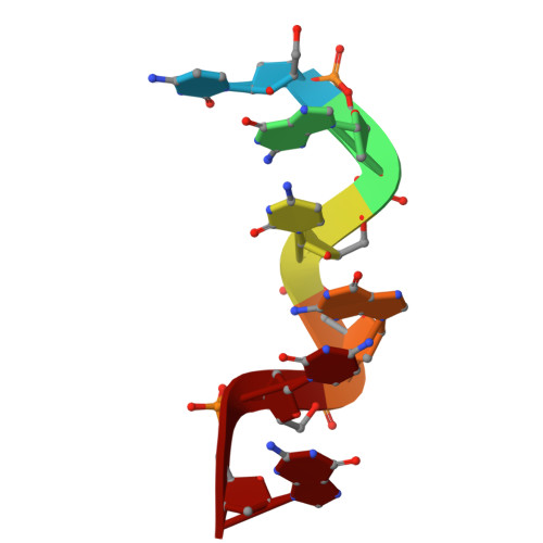
The hydration structure of a Z-DNA hexameric duplex determined by a neutron diffraction technique.

Chatake, T.Tanaka, I.Umino, H.Arai, S.Niimura, N.

(2005) Acta Crystallogr D Biol Crystallogr 61: 1088-1098

  • DOI: https://doi.org/10.1107/S0907444905015581
  • Primary Citation of Related Structures:  
    1V9G, 1WOE

  • PubMed Abstract: 

    In order to reveal the hydration structure of Z-DNA, a neutron diffraction study has been carried out at 1.8 A resolution on a Z-DNA hexamer d(CGCGCG). Neutron diffraction data were collected with the BIX-3 single-crystal diffractometer at the JRR-3 reactor in the Japan Atomic Energy Research Institute (JAERI) using a large crystal (1.6 mm3) obtained from D2O solution. It has been found that almost all the guanine bases have participated in H/D exchange at the C8-H8 group, consistent with the acidic nature of this bond. 44 water molecules were found in the nuclear density maps, of which 29 showed the entire contour of all three atoms (D-O-D). The remaining 15 water molecules had a simple spherical shape, indicating that they were rotationally disordered. An interesting relationship was found between the orientational disorder of the water molecules and their locations. Almost all water molecules in the minor groove were well ordered in the crystal, while 40% of the water molecules in the major groove were rotationally disordered. The hydrogen-bonding networks in the hydration shells have two structural aspects: flexibility and regularity.


  • Organizational Affiliation

    Faculty of Pharmaceutical Sciences, Chiba Institute of Science, Choshi, Chiba 288-0025, Japan.


Macromolecules

Find similar nucleic acids by:  Sequence   |   3D Structure  

Entity ID: 1
MoleculeChains LengthOrganismImage
5'-D(*CP*GP*CP*GP*CP*G)-3'
A, B
6synthetic construct
Sequence Annotations
Expand
  • Reference Sequence
Small Molecules
Ligands 1 Unique
IDChains Name / Formula / InChI Key2D Diagram3D Interactions
SPW
Query on SPW

Download Ideal Coordinates CCD File 
C [auth A]N,N'-BIS(3-AMMONIOPROPYL)BUTANE-1,4-DIAMINIUM
C10 H20 N4
PFNFFQXMRSDOHW-NLAGYVBISA-R
Experimental Data & Validation

Experimental Data

  • Method: NEUTRON DIFFRACTION
  • Resolution: 1.80 Å
  • R-Value Free: 0.294 
  • R-Value Work: 0.222 
  • R-Value Observed: 0.229 
  • Space Group: P 21 21 21
Unit Cell:
Length ( Å )Angle ( ˚ )
a = 18.46α = 90
b = 30.76β = 90
c = 43.18γ = 90
Software Package:
Software NamePurpose
DENZOdata reduction
SCALEPACKdata scaling
CNSrefinement
CNSphasing

Structure Validation

View Full Validation Report


Entry History 

Deposition Data

Revision History  (Full details and data files)

  • Version 1.0: 2005-01-26
    Type: Initial release
  • Version 1.1: 2008-04-27
    Changes: Version format compliance
  • Version 1.2: 2011-07-13
    Changes: Non-polymer description, Version format compliance
  • Version 1.3: 2013-08-21
    Changes: Derived calculations, Other
  • Version 1.4: 2018-03-07
    Changes: Data collection
  • Version 1.5: 2018-06-20
    Changes: Data collection, Experimental preparation
  • Version 1.6: 2018-11-28
    Changes: Data collection, Source and taxonomy
  • Version 1.7: 2018-12-05
    Changes: Data collection
  • Version 1.8: 2023-10-25
    Changes: Data collection, Database references, Derived calculations, Refinement description