1UKR

STRUCTURE OF ENDO-1,4-BETA-XYLANASE C


Experimental Data Snapshot

  • Method: X-RAY DIFFRACTION
  • Resolution: 2.40 Å
  • R-Value Work: 0.179 
  • R-Value Observed: 0.179 

wwPDB Validation   3D Report Full Report


This is version 1.3 of the entry. See complete history


Literature

Three-dimensional structure of Endo-1,4-beta-xylanase I from Aspergillus niger: molecular basis for its low pH optimum.

Krengel, U.Dijkstra, B.W.

(1996) J Mol Biol 263: 70-78

  • DOI: https://doi.org/10.1006/jmbi.1996.0556
  • Primary Citation of Related Structures:  
    1UKR

  • PubMed Abstract: 

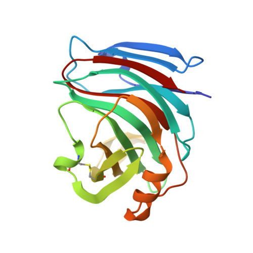
    The crystal structure of endo-1,4-beta-xylanase I from Aspergillus niger has been solved by molecular replacement and was refined to 2.4 A resolution. The final R-factor for all data from 6 to 2.4 A is 17.9%. The A. niger xylanase has a characteristic fold which is unique for family G xylanases (root-mean-square deviation = 1.1 A to Trichoderma reesei xylanase I, which has 53% sequence identity). It consists of a single domain composed predominantly of beta-strands. Two beta-sheets are twisted around a deep, long cleft, which is lined with many aromatic amino acid residues and is large enough to accommodate at least four xylose residues. The two conserved glutamate residues, Glu79 and Glu170, which are likely to be involved in catalysis, reach into this cleft from opposite sides. A niger xylanase I is of particular commercial interest because of its low pH optimum. A model is proposed which explains this low pH optimum compared to other members of xylanase family G.


  • Organizational Affiliation

    BIOSON Research Institute, University of Groningen, The Netherlands.


Macromolecules
Find similar proteins by:  (by identity cutoff)  |  3D Structure
Entity ID: 1
MoleculeChains Sequence LengthOrganismDetailsImage
ENDO-1,4-B-XYLANASE I
A, B, C, D
184Aspergillus nigerMutation(s): 0 
EC: 3.2.1.8
UniProt
Find proteins for P55329 (Aspergillus niger)
Explore P55329 
Go to UniProtKB:  P55329
Entity Groups  
Sequence Clusters30% Identity50% Identity70% Identity90% Identity95% Identity100% Identity
UniProt GroupP55329
Sequence Annotations
Expand
  • Reference Sequence
Experimental Data & Validation

Experimental Data

  • Method: X-RAY DIFFRACTION
  • Resolution: 2.40 Å
  • R-Value Work: 0.179 
  • R-Value Observed: 0.179 
  • Space Group: P 21 21 21
Unit Cell:
Length ( Å )Angle ( ˚ )
a = 84.3α = 90
b = 85.3β = 90
c = 113.8γ = 90
Software Package:
Software NamePurpose
XDSdata scaling
BRUTEmodel building
X-PLORrefinement
XDSdata reduction
BRUTEphasing

Structure Validation

View Full Validation Report


Entry History 

Deposition Data

Revision History  (Full details and data files)

  • Version 1.0: 1997-12-24
    Type: Initial release
  • Version 1.1: 2008-03-24
    Changes: Version format compliance
  • Version 1.2: 2011-07-13
    Changes: Version format compliance
  • Version 1.3: 2023-08-09
    Changes: Data collection, Database references, Refinement description