Compared structures of the free nicotinic acetylcholine receptor main immunogenic region (MIR) decapeptide and the antibody-bound [A76]MIR analogue: a molecular dynamics simulation from two-dimensional NMR data.
Orlewski, P., Marraud, M., Cung, M.T., Tsikaris, V., Sakarellos-Daitsiotis, M., Sakarellos, C., Vatzaki, E., Tzartos, S.J.(1996) Biopolymers 40: 419-432
- PubMed: 9062066
- DOI: https://doi.org/10.1002/(sici)1097-0282(1996)40:5<419::aid-bip1>3.0.co;2-z
- Primary Citation of Related Structures:
1TOR, 1TOS - PubMed Abstract:
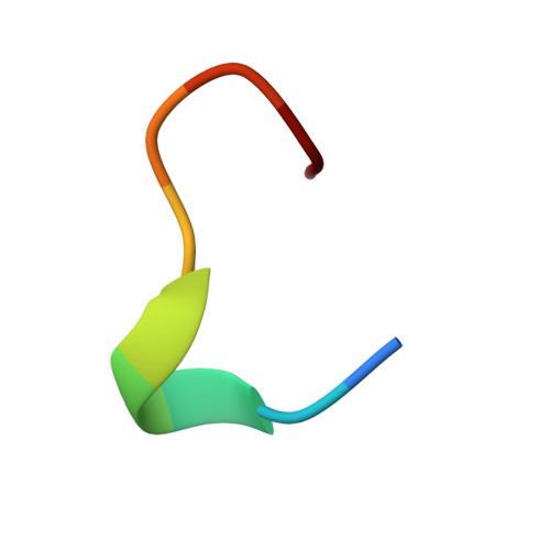
Monoclonal antibodies against the main immunogenic region (MIR) of the muscle acetylcholine receptor (AChR) are capable of inducing experimental myasthenia gravis (MG) in animals. The epitope of these antibodies has been localized between residues 67 and 76 of the AChR alpha-subunit. The conformation in solution of the Torpedo californica MIR peptide and of its [A76] MIR analogue have been analyzed using molecular modeling based on nmr interproton distances and J-derived phi dihedral angles. Molecular dynamics simulations including dimethyl-sulfoxide as explicit solvent have been carried out on the free MIR peptide. Calculation of the structure of the [A76]MIR analogue bound to an anti-MIR monoclonal antibody have been performed in the presence of water molecules. A tightly folded structure appears for both peptides with alpha beta-folded N-terminal N68-P-A-D71 sequence of type I in the free state and type III in the mAb6-bound state. The C-terminal sequence is folded in two different ways according to the result in the free and bound state of the peptides: two overlapping beta/beta or beta/alpha turns result in a short helical sequence in the free MIR peptide, whereas the bound analogue is folded by uncommon hydrogen bond closing an 11-membered cycle. This structural evolution is essentially the result of the reorientation of the hydrophobic side chains that are probably directly involved in peptide--antibody recognition.
Organizational Affiliation:
Laboratoire de Chimie-Physique Macromoléculaire CNRS-URA 494 ENSIC-INPL, Nancy, France.