1T3N

Structure of the catalytic core of DNA polymerase Iota in complex with DNA and dTTP


Experimental Data Snapshot

  • Method: X-RAY DIFFRACTION
  • Resolution: 2.30 Å
  • R-Value Free: 0.286 
  • R-Value Work: 0.265 

wwPDB Validation   3D Report Full Report


Ligand Structure Quality Assessment 


This is version 1.4 of the entry. See complete history

Re-refinement Note

A newer entry is available that reflects an alternative modeling of the original data: 1ZET


Literature

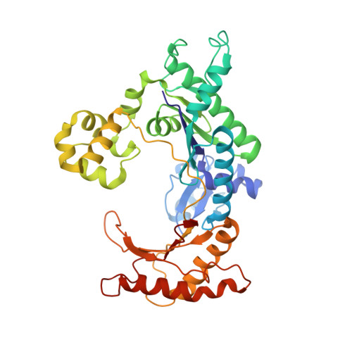
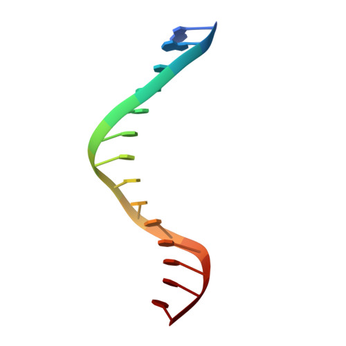
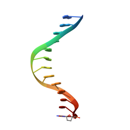
Replication by human DNA polymerase-iota occurs by Hoogsteen base-pairing.

Nair, D.T.Johnson, R.E.Prakash, S.Prakash, L.Aggarwal, A.K.

(2004) Nature 430: 377-380

  • DOI: https://doi.org/10.1038/nature02692
  • Primary Citation of Related Structures:  
    1T3N

  • PubMed Abstract: 

    Almost all DNA polymerases show a strong preference for incorporating the nucleotide that forms the correct Watson-Crick base pair with the template base. In addition, the catalytic efficiencies with which any given polymerase forms the four possible correct base pairs are roughly the same. Human DNA polymerase-iota (hPoliota), a member of the Y family of DNA polymerases, is an exception to these rules. hPoliota incorporates the correct nucleotide opposite a template adenine with a several hundred to several thousand fold greater efficiency than it incorporates the correct nucleotide opposite a template thymine, whereas its efficiency for correct nucleotide incorporation opposite a template guanine or cytosine is intermediate between these two extremes. Here we present the crystal structure of hPoliota bound to a template primer and an incoming nucleotide. The structure reveals a polymerase that is 'specialized' for Hoogsteen base-pairing, whereby the templating base is driven to the syn conformation. Hoogsteen base-pairing offers a basis for the varied efficiencies and fidelities of hPoliota opposite different template bases, and it provides an elegant mechanism for promoting replication through minor-groove purine adducts that interfere with replication.


  • Organizational Affiliation

    Structural Biology Program, Department of Physiology and Biophysics, Mount Sinai School of Medicine, Box 1677, 1425 Madison Avenue, New York, New York 10029, USA.


Macromolecules

Find similar proteins by:  (by identity cutoff)  |  3D Structure
Entity ID: 3
MoleculeChains Sequence LengthOrganismDetailsImage
polymerase (DNA directed) iotaC [auth A],
D [auth B]
388Homo sapiensMutation(s): 0 
Gene Names: POLI
UniProt & NIH Common Fund Data Resources
Find proteins for Q9UNA4 (Homo sapiens)
Explore Q9UNA4 
Go to UniProtKB:  Q9UNA4
PHAROS:  Q9UNA4
GTEx:  ENSG00000101751 
Entity Groups  
Sequence Clusters30% Identity50% Identity70% Identity90% Identity95% Identity100% Identity
UniProt GroupQ9UNA4
Sequence Annotations
Expand
  • Reference Sequence

Find similar nucleic acids by:  Sequence   |   3D Structure  

Entity ID: 1
MoleculeChains LengthOrganismImage
Template DNA strandA [auth T]14N/A
Sequence Annotations
Expand
  • Reference Sequence

Find similar nucleic acids by:  Sequence   |   3D Structure  

Entity ID: 2
MoleculeChains LengthOrganismImage
Primer DNA strandB [auth P]13N/A
Sequence Annotations
Expand
  • Reference Sequence
Small Molecules
Ligands 2 Unique
IDChains Name / Formula / InChI Key2D Diagram3D Interactions
TTP
Query on TTP

Download Ideal Coordinates CCD File 
F [auth B]THYMIDINE-5'-TRIPHOSPHATE
C10 H17 N2 O14 P3
NHVNXKFIZYSCEB-XLPZGREQSA-N
MG
Query on MG

Download Ideal Coordinates CCD File 
E [auth B]MAGNESIUM ION
Mg
JLVVSXFLKOJNIY-UHFFFAOYSA-N
Experimental Data & Validation

Experimental Data

  • Method: X-RAY DIFFRACTION
  • Resolution: 2.30 Å
  • R-Value Free: 0.286 
  • R-Value Work: 0.265 
  • Space Group: P 65
Unit Cell:
Length ( Å )Angle ( ˚ )
a = 98.831α = 90
b = 98.831β = 90
c = 202.755γ = 120
Software Package:
Software NamePurpose
HKL-2000data collection
SCALEPACKdata scaling
SOLVEphasing
CNSrefinement
HKL-2000data reduction

Structure Validation

View Full Validation Report


Ligand Structure Quality Assessment 


Entry History 

Deposition Data

Revision History  (Full details and data files)

  • Version 1.0: 2004-07-20
    Type: Initial release
  • Version 1.1: 2008-04-30
    Changes: Version format compliance
  • Version 1.2: 2011-07-13
    Changes: Version format compliance
  • Version 1.3: 2015-09-09
    Changes: Other
  • Version 1.4: 2018-04-18
    Changes: Data collection, Derived calculations