1SON

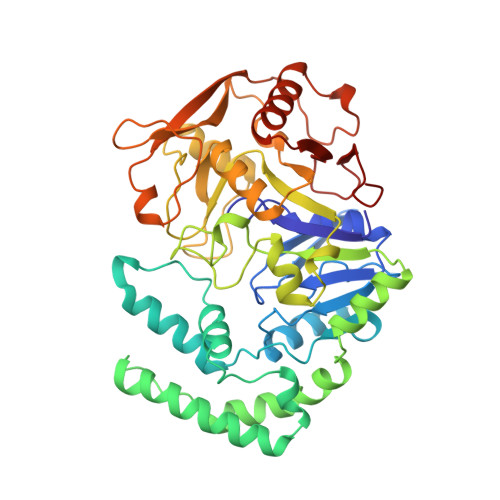
ADENYLOSUCCINATE SYNTHETASE IN COMPLEX WITH THE NATURAL FEEDBACK INHIBITOR AMP


Experimental Data Snapshot

  • Method: X-RAY DIFFRACTION
  • Resolution: 2.55 Å
  • R-Value Work: 0.187 
  • R-Value Observed: 0.187 

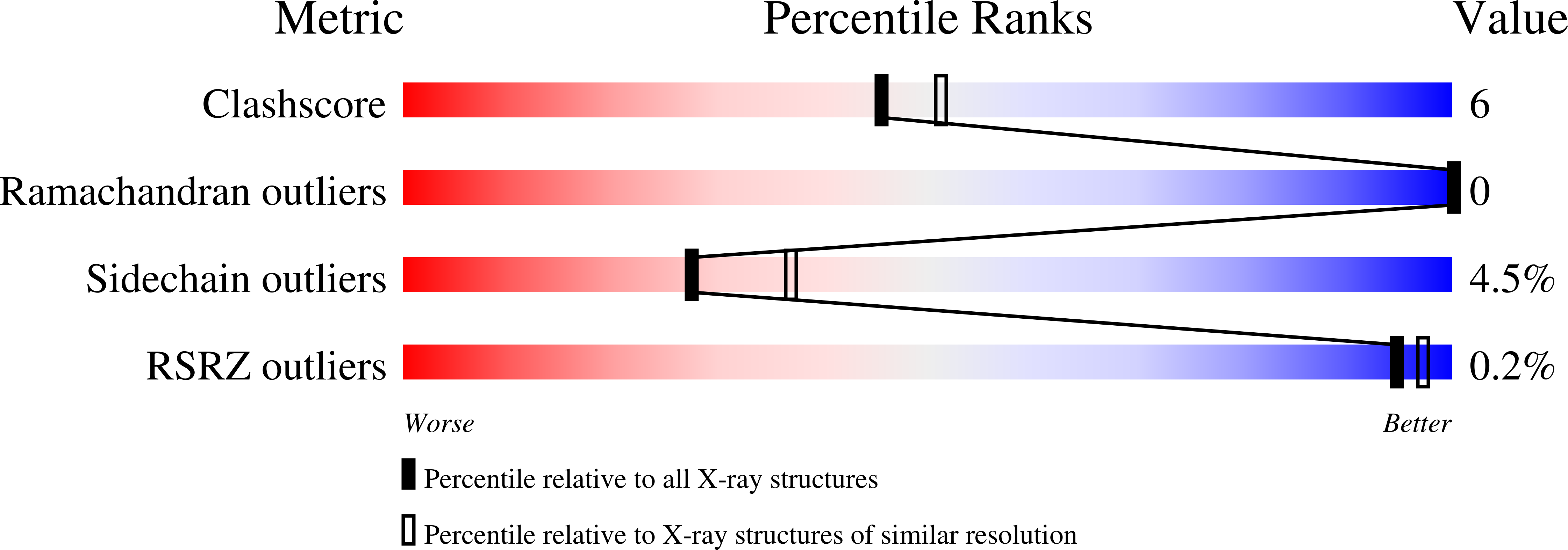
wwPDB Validation   3D Report Full Report


Ligand Structure Quality Assessment 


This is version 1.4 of the entry. See complete history


Literature

The mode of action and the structure of a herbicide in complex with its target: binding of activated hydantocidin to the feedback regulation site of adenylosuccinate synthetase.

Fonne-Pfister, R.Chemla, P.Ward, E.Girardet, M.Kreuz, K.E.Honzatko, R.B.Fromm, H.J.Schar, H.P.Grutter, M.G.Cowan-Jacob, S.W.

(1996) Proc Natl Acad Sci U S A 93: 9431-9436

  • DOI: https://doi.org/10.1073/pnas.93.18.9431
  • Primary Citation of Related Structures:  
    1SON, 1SOO

  • PubMed Abstract: 

    (+)-Hydantocidin, a recently discovered natural spironucleoside with potent herbicidal activity, is shown to be a proherbicide that, after phosphorylation at the 5' position, inhibits adenylosuccinate synthetase, an enzyme involved in de novo purine synthesis. The mode of binding of hydantocidin 5'-monophosphate to the target enzyme was analyzed by determining the crystal structure of the enzyme-inhibitor complex at 2.6-A resolution. It was found that adenylosuccinate synthetase binds the phosphorylated compound in the same fashion as it does adenosine 5'-monophosphate, the natural feedback regulator of this enzyme. This work provides the first crystal structure of a herbicide-target complex reported to date.


  • Organizational Affiliation

    Pharmaceutical Division, Ciba-Geigy Ltd., Basel, Switzerland.


Macromolecules
Find similar proteins by:  (by identity cutoff)  |  3D Structure
Entity ID: 1
MoleculeChains Sequence LengthOrganismDetailsImage
ADENYLOSUCCINATE SYNTHETASE431Escherichia coliMutation(s): 0 
EC: 6.3.4.4
UniProt
Find proteins for P0A7D4 (Escherichia coli (strain K12))
Explore P0A7D4 
Go to UniProtKB:  P0A7D4
Entity Groups  
Sequence Clusters30% Identity50% Identity70% Identity90% Identity95% Identity100% Identity
UniProt GroupP0A7D4
Sequence Annotations
Expand
  • Reference Sequence
Small Molecules
Ligands 2 Unique
IDChains Name / Formula / InChI Key2D Diagram3D Interactions
AMP
Query on AMP

Download Ideal Coordinates CCD File 
B [auth A]ADENOSINE MONOPHOSPHATE
C10 H14 N5 O7 P
UDMBCSSLTHHNCD-KQYNXXCUSA-N
BME
Query on BME

Download Ideal Coordinates CCD File 
C [auth A]BETA-MERCAPTOETHANOL
C2 H6 O S
DGVVWUTYPXICAM-UHFFFAOYSA-N
Experimental Data & Validation

Experimental Data

  • Method: X-RAY DIFFRACTION
  • Resolution: 2.55 Å
  • R-Value Work: 0.187 
  • R-Value Observed: 0.187 
  • Space Group: P 43 21 2
Unit Cell:
Length ( Å )Angle ( ˚ )
a = 70.5α = 90
b = 70.5β = 90
c = 201.2γ = 90
Software Package:
Software NamePurpose
X-PLORmodel building
X-PLORrefinement
XDSdata reduction
CCP4data scaling
ROTAVATAdata scaling
X-PLORphasing

Structure Validation

View Full Validation Report


Ligand Structure Quality Assessment 


Entry History 

Deposition Data

Revision History  (Full details and data files)

  • Version 1.0: 1997-09-04
    Type: Initial release
  • Version 1.1: 2008-03-24
    Changes: Version format compliance
  • Version 1.2: 2011-07-13
    Changes: Derived calculations, Version format compliance
  • Version 1.3: 2011-11-16
    Changes: Atomic model
  • Version 1.4: 2023-08-09
    Changes: Database references, Derived calculations, Other, Refinement description