1RTX

Crystal Structure of Synechocystis Hemoglobin with a Covalent Heme Linkage


Experimental Data Snapshot

  • Method: X-RAY DIFFRACTION
  • Resolution: 1.80 Å
  • R-Value Free: 0.224 
  • R-Value Work: 0.218 
  • R-Value Observed: 0.218 

wwPDB Validation   3D Report Full Report


Ligand Structure Quality Assessment 


This is version 1.3 of the entry. See complete history


Literature

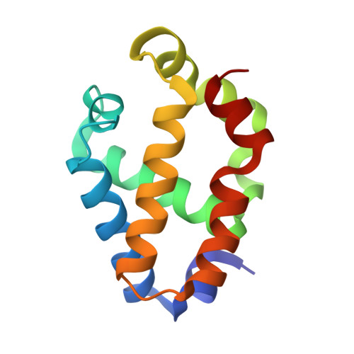
The crystal structure of synechocystis hemoglobin with a covalent heme linkage.

Hoy, J.A.Kundu, S.Trent, J.T.Ramaswamy, S.Hargrove, M.S.

(2004) J Biol Chem 279: 16535-16542

  • DOI: https://doi.org/10.1074/jbc.M313707200
  • Primary Citation of Related Structures:  
    1RTX

  • PubMed Abstract: 

    The x-ray crystal structure of Synechocystis hemoglobin has been solved to a resolution of 1.8 A. The conformation of this structure is surprisingly different from that of the previously reported solution structure, probably due in part to a covalent linkage between the heme 2-vinyl and His117 that is present in the crystal structure but not in the structure solved by NMR. Synechocystis hemoglobin is a hexacoordinate hemoglobin in which the heme iron is coordinated by both the proximal and distal histidines. It is also a member of the "truncated hemoglobin" family that is much shorter in primary structure than vertebrate and plant hemoglobins. In contrast to other truncated hemoglobins, the crystal structure of Synechocystis hemoglobin displays no "ligand tunnel" and shows that several important amino acid side chains extrude into the solvent instead of residing inside the heme pocket. The stereochemistry of hexacoordination is compared with other hexacoordinate hemoglobins and cytochromes in an effort to illuminate factors contributing to ligand affinity in hexacoordinate hemoglobins.


  • Organizational Affiliation

    Department of Biochemistry, Biophysics, and Molecular Biology, Iowa State University, Ames, Iowa 50011, USA.


Macromolecules
Find similar proteins by:  (by identity cutoff)  |  3D Structure
Entity ID: 1
MoleculeChains Sequence LengthOrganismDetailsImage
Cyanoglobin123Synechocystis sp. PCC 6803Mutation(s): 0 
Gene Names: GLBNSLR2097
UniProt
Find proteins for P73925 (Synechocystis sp. (strain PCC 6803 / Kazusa))
Explore P73925 
Go to UniProtKB:  P73925
Entity Groups  
Sequence Clusters30% Identity50% Identity70% Identity90% Identity95% Identity100% Identity
UniProt GroupP73925
Sequence Annotations
Expand
  • Reference Sequence
Experimental Data & Validation

Experimental Data

  • Method: X-RAY DIFFRACTION
  • Resolution: 1.80 Å
  • R-Value Free: 0.224 
  • R-Value Work: 0.218 
  • R-Value Observed: 0.218 
  • Space Group: P 21 21 21
Unit Cell:
Length ( Å )Angle ( ˚ )
a = 42.5α = 90
b = 46β = 90
c = 61γ = 90
Software Package:
Software NamePurpose
d*TREKdata scaling
d*TREKdata reduction
REFMACrefinement
SOLVEphasing
RESOLVEphasing

Structure Validation

View Full Validation Report


Ligand Structure Quality Assessment 


Entry History 

Deposition Data

Revision History  (Full details and data files)

  • Version 1.0: 2004-04-20
    Type: Initial release
  • Version 1.1: 2008-04-29
    Changes: Version format compliance
  • Version 1.2: 2011-07-13
    Changes: Source and taxonomy, Version format compliance
  • Version 1.3: 2024-02-14
    Changes: Data collection, Database references, Derived calculations