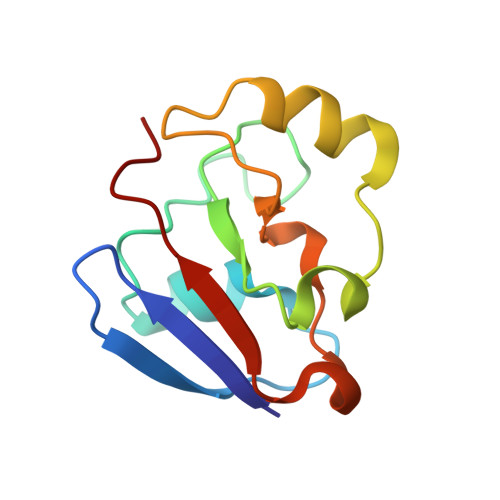
Structure of C73G putidaredoxin from Pseudomonas putida.
Smith, N., Mayhew, M., Holden, M.J., Kelly, H., Robinson, H., Heroux, A., Vilker, V.L., Gallagher, D.T.(2004) Acta Crystallogr D Biol Crystallogr 60: 816-822
- PubMed: 15103126
- DOI: https://doi.org/10.1107/S0907444904003348
- Primary Citation of Related Structures:
1R7S - PubMed Abstract:
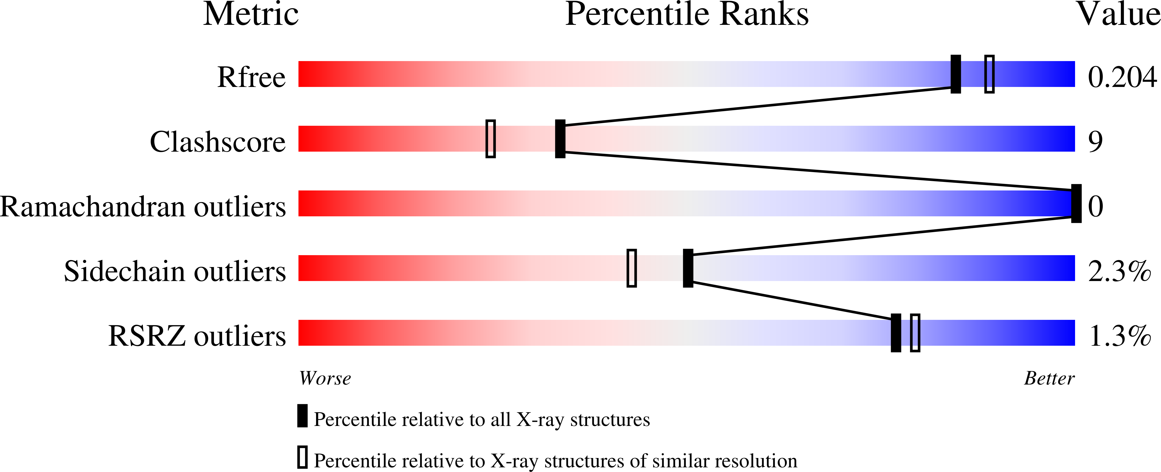
The structure of the C73G mutant of putidaredoxin (Pdx), the Fe(2)S(2) ferredoxin that supplies electrons to cytochrome CYP101 (p450cam) for camphor oxidation, is reported at 1.9 A resolution in a C2 crystal form. The structure was solved by single-wavelength iron anomalous diffraction, which yielded electron density above the 2sigma level for over 97% of the non-H atoms in the protein. The final structure with R = 0.19 and R(free) = 0.21 has been deposited in the Protein Data Bank with accession code 1r7s. The C2 crystal contains three Pdx molecules in the asymmetric unit, giving three independent models of the protein that are very similar (r.m.s.d. < 0.3 A for the 106 C(alpha) atoms). The unusually high solvent fraction of 80% results in comparatively few crystal-packing artifacts. The structure is briefly compared with the recently reported crystal structures of the C73S and C73S/C85S mutants. In general, the eight independent molecules in the three crystal structures (three in C73G, three in C73S and two in C73S/C85S) are much more similar to each other than to the previously reported NMR structure of wild-type Pdx in solution. The present findings show a unanimous structure in some regions crucial for electron-transfer interactions, including the cluster-binding loop 39-48 and the cytochrome-interaction region of Asp38 and Trp106. In addition, the Cys45 amide group donates a hydrogen bond to cluster sulfur S1, with Ala46 adopting an Lalpha conformation, in all three molecules in the crystal.
Organizational Affiliation:
Biotechnology Division of the National Institute of Standards and Technology, Gaithersburg, MD 20899-8312, USA.