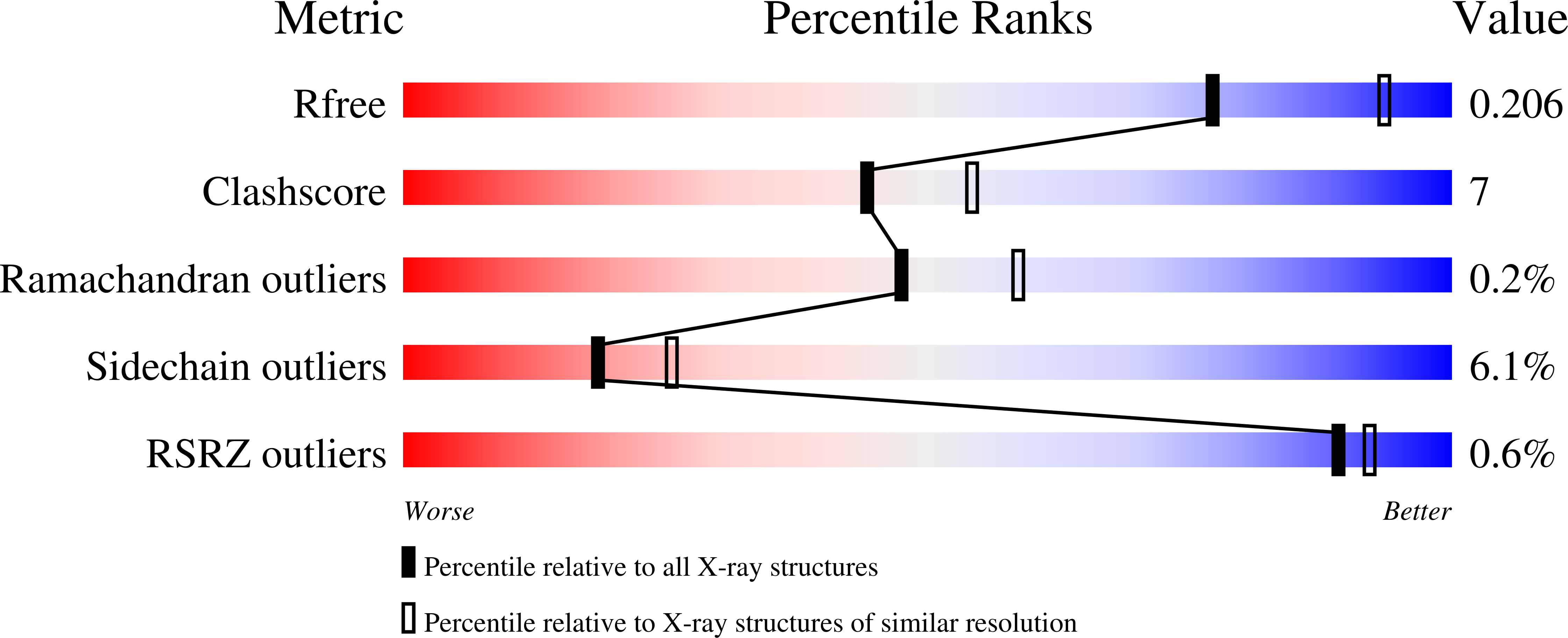
1R56

UNCOMPLEXED URATE OXIDASE FROM ASPERGILLUS FLAVUS


Experimental Data Snapshot

  • Method: X-RAY DIFFRACTION
  • Resolution: 2.30 Å
  • R-Value Free: 0.214 
  • R-Value Work: 0.162 

wwPDB Validation   3D Report Full Report


This is version 1.4 of the entry. See complete history


Literature

Complexed and ligand-free high-resolution structures of urate oxidase (Uox) from Aspergillus flavus: a reassignment of the active-site binding mode.

Retailleau, P.Colloc'h, N.Vivares, D.Bonnete, F.Castro, B.El-Hajji, M.Mornon, J.P.Monard, G.Prange, T.

(2004) Acta Crystallogr D Biol Crystallogr 60: 453-462

  • DOI: https://doi.org/10.1107/S0907444903029718
  • Primary Citation of Related Structures:  
    1R4S, 1R4U, 1R51, 1R56

  • PubMed Abstract: 

    High-resolution X-ray structures of the complexes of Aspergillus flavus urate oxidase (Uox) with three inhibitors, 8-azaxanthin (AZA), 9-methyl uric acid (MUA) and oxonic acid (OXC), were determined in an orthorhombic space group (I222). In addition, the ligand-free enzyme was also crystallized in a monoclinic form (P2(1)) and its structure determined. Higher accuracy in the three new enzyme-inhibitor complex structures (Uox-AZA, Uox-MUA and Uox-OXC) with respect to the previously determined structure of Uox-AZA (PDB code 1uox) leads to a reversed position of the inhibitor in the active site of the enzyme. The corrected anchoring of the substrate (uric acid) allows an improvement in the understanding of the enzymatic mechanism of urate oxidase.


  • Organizational Affiliation

    LURE, Centre Universitaire Paris-Sud, Bâtiment 209D, BP 34, 91898 Orsay CEDEX, France. retailleau@lure.u-psud.fr


Macromolecules
Find similar proteins by:  (by identity cutoff)  |  3D Structure
Entity ID: 1
MoleculeChains Sequence LengthOrganismDetailsImage
Uricase
A, B, C, D, E
A, B, C, D, E, F, G, H
301Aspergillus flavusMutation(s): 1 
EC: 1.7.3.3
UniProt
Find proteins for Q00511 (Aspergillus flavus)
Explore Q00511 
Go to UniProtKB:  Q00511
Entity Groups  
Sequence Clusters30% Identity50% Identity70% Identity90% Identity95% Identity100% Identity
UniProt GroupQ00511
Sequence Annotations
Expand
  • Reference Sequence
Small Molecules
Modified Residues  1 Unique
IDChains TypeFormula2D DiagramParent
SAC
Query on SAC
A, B, C, D, E
A, B, C, D, E, F, G, H
L-PEPTIDE LINKINGC5 H9 N O4SER
Experimental Data & Validation

Experimental Data

  • Method: X-RAY DIFFRACTION
  • Resolution: 2.30 Å
  • R-Value Free: 0.214 
  • R-Value Work: 0.162 
  • Space Group: P 1 21 1
Unit Cell:
Length ( Å )Angle ( ˚ )
a = 82.59α = 90
b = 141.843β = 92.69
c = 134.959γ = 90
Software Package:
Software NamePurpose
DENZOdata reduction
SCALEPACKdata scaling
BUSTER-TNTrefinement
EPMRphasing

Structure Validation

View Full Validation Report


Entry History 

Deposition Data

Revision History  (Full details and data files)

  • Version 1.0: 2004-03-02
    Type: Initial release
  • Version 1.1: 2008-04-29
    Changes: Version format compliance
  • Version 1.2: 2011-07-13
    Changes: Version format compliance
  • Version 1.3: 2018-02-28
    Changes: Database references, Structure summary
  • Version 1.4: 2023-08-23
    Changes: Data collection, Database references, Derived calculations, Refinement description