1QRP

Human pepsin 3A in complex with a phosphonate inhibitor IVA-VAL-VAL-LEU(P)-(O)PHE-ALA-ALA-OME


Experimental Data Snapshot

  • Method: X-RAY DIFFRACTION
  • Resolution: 1.96 Å
  • R-Value Observed: 0.200 

wwPDB Validation   3D Report Full Report


Ligand Structure Quality Assessment 


This is version 1.3 of the entry. See complete history


Literature

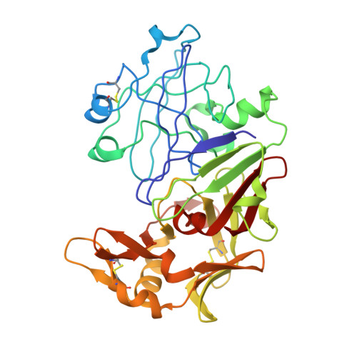
Structural study of the complex between human pepsin and a phosphorus-containing peptidic -transition-state analog.

Fujinaga, M.Cherney, M.M.Tarasova, N.I.Bartlett, P.A.Hanson, J.E.James, M.N.

(2000) Acta Crystallogr D Biol Crystallogr 56: 272-279

  • DOI: https://doi.org/10.1107/s0907444999016376
  • Primary Citation of Related Structures:  
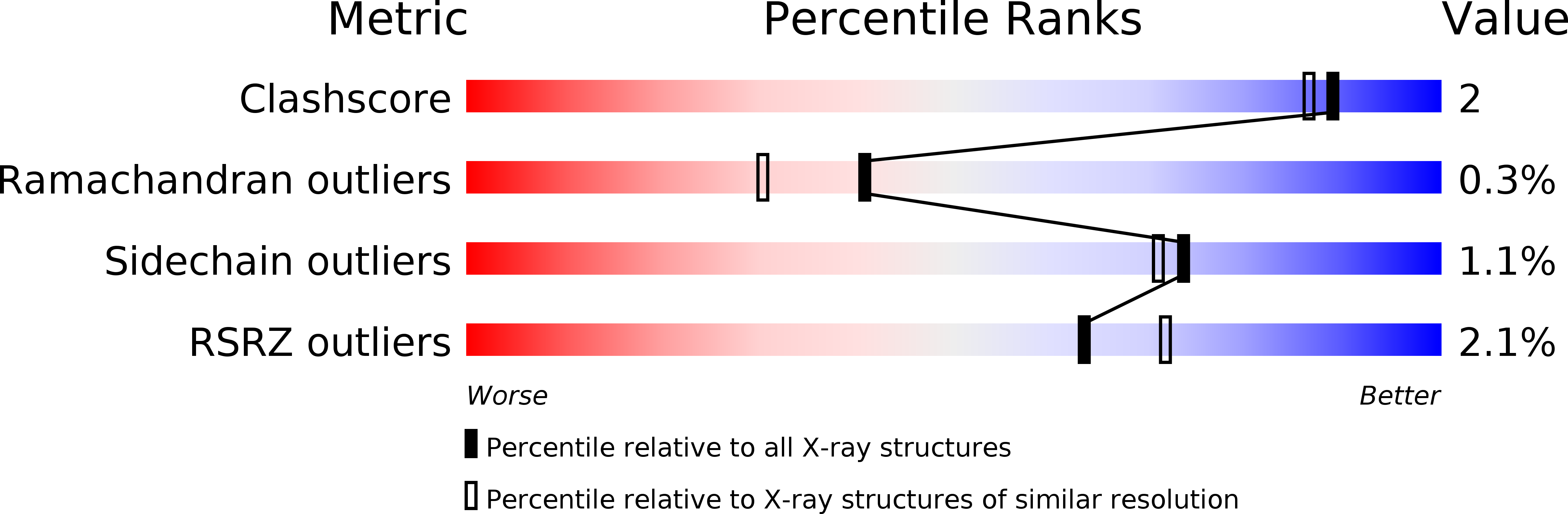
    1QRP

  • PubMed Abstract: 

    The refined crystal structure of the complex between human pepsin and a synthetic phosphonate inhibitor, Iva-Val-Val-Leu(P)-(O)Phe-Ala-Ala-OMe [Iva = isovaleryl, Leu(P) = the phosphinic acid analog of L-leucine, (O)Phe = L-3-phenyllactic acid, OMe = methyl ester], is presented. The structure was refined using diffraction data between 30 and 1.96 A resolution to a final R factor ( summation operator| |F(o)| - |F(c)| | / summation operator|F(o)|, where |F(o)| and |F(c)| are the observed and calculated structure-factor amplitudes, respectively) of 20.0%. The interactions of the inhibitor with the enzyme show the locations of the binding sites on the enzyme from S4 to S3'. Modeling of the inhibitor binding to porcine pepsin shows very similar binding sites, except at S4. Comparison of the complex structure with the structures of related inhibitors bound to penicillopepsin helps to rationalize the observed differences in the binding constants. The convergence of reaction mechanisms and geometries in different families of proteinases is also discussed.


  • Organizational Affiliation

    Medical Research Council of Canada Group in Protein Structure and Function, Department of Biochemistry, University of Alberta, Edmonton, Alberta T6G 2H7, Canada.


Macromolecules
Find similar proteins by:  (by identity cutoff)  |  3D Structure
Entity ID: 1
MoleculeChains Sequence LengthOrganismDetailsImage
PEPSIN 3AA [auth E]326Homo sapiensMutation(s): 0 
UniProt & NIH Common Fund Data Resources
Find proteins for P0DJD7 (Homo sapiens)
Explore P0DJD7 
Go to UniProtKB:  P0DJD7
PHAROS:  P0DJD7
GTEx:  ENSG00000229183 
Entity Groups  
Sequence Clusters30% Identity50% Identity70% Identity90% Identity95% Identity100% Identity
UniProt GroupP0DJD7
Sequence Annotations
Expand
  • Reference Sequence
Small Molecules
Ligands 1 Unique
IDChains Name / Formula / InChI Key2D Diagram3D Interactions
HH0
Query on HH0

Download Ideal Coordinates CCD File 
B [auth E]methyl N-[(2S)-2-({(S)-hydroxy[(1R)-3-methyl-1-{[N-(3-methylbutanoyl)-L-valyl-L-valyl]amino}butyl]phosphoryl}oxy)-3-phenylpropanoyl]-L-alanyl-L-alaninate
C36 H60 N5 O10 P
CEPZPGMVMASQKU-ORDNGIEBSA-N
Biologically Interesting Molecules (External Reference) 1 Unique
Experimental Data & Validation

Experimental Data

  • Method: X-RAY DIFFRACTION
  • Resolution: 1.96 Å
  • R-Value Observed: 0.200 
  • Space Group: P 21 21 21
Unit Cell:
Length ( Å )Angle ( ˚ )
a = 71.9α = 90
b = 151.27β = 90
c = 40.79γ = 90
Software Package:
Software NamePurpose
SDMSdata collection
SDMSdata reduction
XTALVIEWrefinement
GROMOS87refinement
SDMSdata scaling

Structure Validation

View Full Validation Report


Ligand Structure Quality Assessment 


Entry History 

Deposition Data

Revision History  (Full details and data files)

  • Version 1.0: 1999-06-18
    Type: Initial release
  • Version 1.1: 2008-04-27
    Changes: Version format compliance
  • Version 1.2: 2011-07-13
    Changes: Atomic model, Database references, Derived calculations, Non-polymer description, Structure summary, Version format compliance
  • Version 1.3: 2012-12-12
    Changes: Other