1PX8

Crystal structure of beta-D-xylosidase from Thermoanaerobacterium saccharolyticum, a family 39 glycoside hydrolase


Experimental Data Snapshot

  • Method: X-RAY DIFFRACTION
  • Resolution: 2.40 Å
  • R-Value Free: 0.271 
  • R-Value Work: 0.224 
  • R-Value Observed: 0.224 

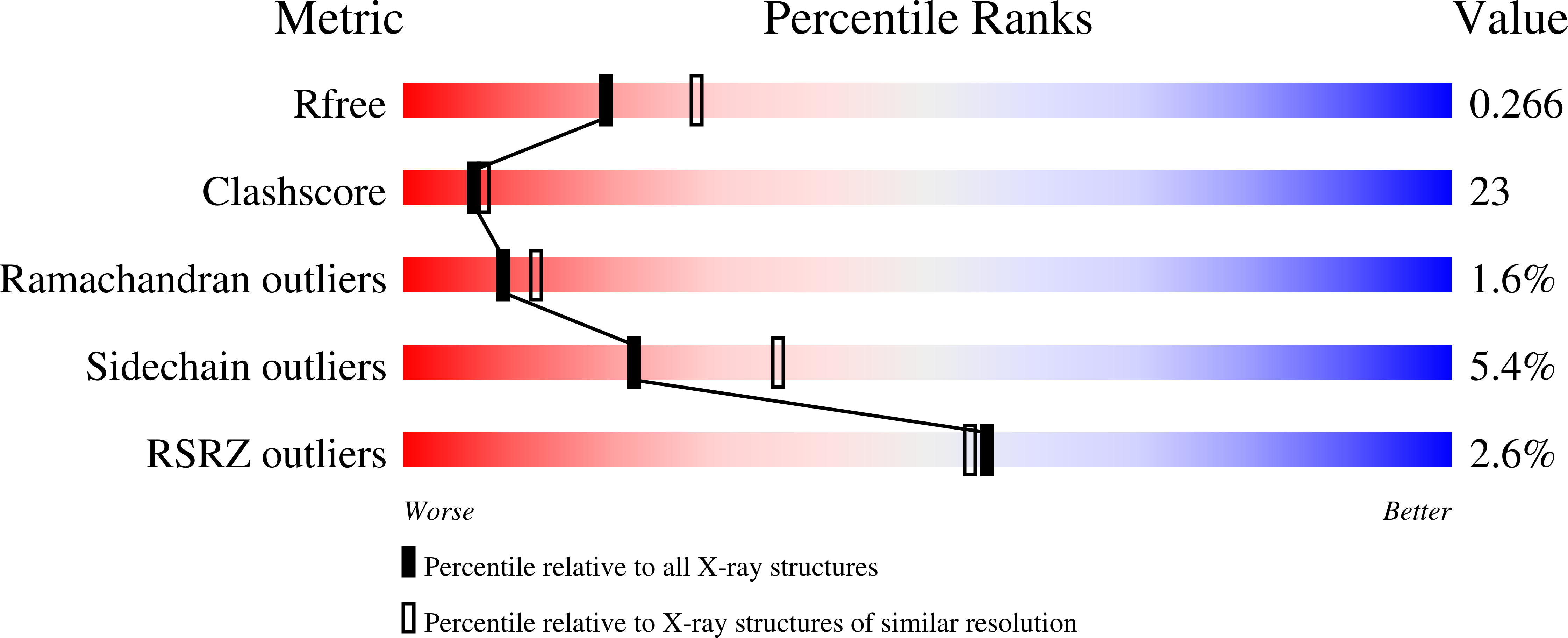
wwPDB Validation   3D Report Full Report


Ligand Structure Quality Assessment 


This is version 2.1 of the entry. See complete history


Literature

Crystal structure of beta-D-xylosidase from Thermoanaerobacterium saccharolyticum, a family 39 glycoside hydrolase.

Yang, J.K.Yoon, H.J.Ahn, H.J.Lee, B.I.Pedelacq, J.D.Liong, E.C.Berendzen, J.Laivenieks, M.Vieille, C.Zeikus, G.J.Vocadlo, D.J.Withers, S.G.Suh, S.W.

(2004) J Mol Biol 335: 155-165

  • DOI: https://doi.org/10.1016/j.jmb.2003.10.026
  • Primary Citation of Related Structures:  
    1PX8, 1UHV

  • PubMed Abstract: 

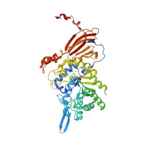
    1,4-beta-D-Xylan is the major component of plant cell-wall hemicelluloses. beta-D-Xylosidases are involved in the breakdown of xylans into xylose and belong to families 3, 39, 43, 52, and 54 of glycoside hydrolases. Here, we report the first crystal structure of a member of family 39 glycoside hydrolase, i.e. beta-D-xylosidase from Thermoanaerobacterium saccharolyticum strain B6A-RI. This study also represents the first structure of any beta-xylosidase of the above five glycoside hydrolase families. Each monomer of T. saccharolyticum beta-xylosidase comprises three distinct domains; a catalytic domain of the canonical (beta/alpha)(8)-barrel fold, a beta-sandwich domain, and a small alpha-helical domain. We have determined the structure in two forms: D-xylose-bound enzyme and a covalent 2-deoxy-2-fluoro-alpha-D-xylosyl-enzyme intermediate complex, thus providing two snapshots in the reaction pathway. This study provides structural evidence for the proposed double displacement mechanism that involves a covalent intermediate. Furthermore, it reveals possible functional roles for His228 as the auxiliary acid/base and Glu323 as a key residue in substrate recognition.


  • Organizational Affiliation

    Structural Proteomics Laboratory, Department of Chemistry, College of Natural Sciences, Seoul National University, 151-742, Seoul, South Korea.


Macromolecules
Find similar proteins by:  (by identity cutoff)  |  3D Structure
Entity ID: 1
MoleculeChains Sequence LengthOrganismDetailsImage
Beta-xylosidase
A, B
500Thermoanaerobacterium saccharolyticumMutation(s): 0 
EC: 3.2.1.37
UniProt
Find proteins for P36906 (Thermoanaerobacterium saccharolyticum)
Explore P36906 
Go to UniProtKB:  P36906
Entity Groups  
Sequence Clusters30% Identity50% Identity70% Identity90% Identity95% Identity100% Identity
UniProt GroupP36906
Sequence Annotations
Expand
  • Reference Sequence
Small Molecules
Experimental Data & Validation

Experimental Data

  • Method: X-RAY DIFFRACTION
  • Resolution: 2.40 Å
  • R-Value Free: 0.271 
  • R-Value Work: 0.224 
  • R-Value Observed: 0.224 
  • Space Group: P 43 21 2
Unit Cell:
Length ( Å )Angle ( ˚ )
a = 92.719α = 90
b = 92.719β = 90
c = 241.296γ = 90
Software Package:
Software NamePurpose
CNSrefinement
DENZOdata reduction
SCALEPACKdata scaling
SOLVEphasing

Structure Validation

View Full Validation Report


Ligand Structure Quality Assessment 


Entry History 

Revision History  (Full details and data files)

  • Version 1.0: 2003-12-23
    Type: Initial release
  • Version 1.1: 2008-04-29
    Changes: Version format compliance
  • Version 1.2: 2011-07-13
    Changes: Derived calculations, Version format compliance
  • Version 2.0: 2020-07-29
    Type: Remediation
    Reason: Carbohydrate remediation
    Changes: Atomic model, Data collection, Derived calculations, Structure summary
  • Version 2.1: 2024-03-13
    Changes: Data collection, Database references, Structure summary