1P9N

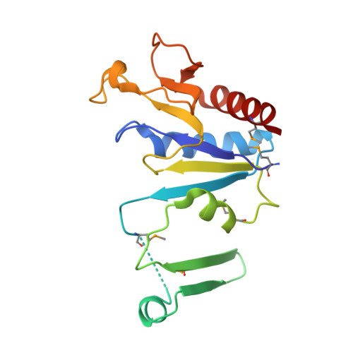
Crystal structure of Escherichia coli MobB.


Experimental Data Snapshot

  • Method: X-RAY DIFFRACTION
  • Resolution: 2.80 Å
  • R-Value Free: 0.308 
  • R-Value Work: 0.275 
  • R-Value Observed: 0.275 

wwPDB Validation   3D Report Full Report


This is version 1.3 of the entry. See complete history


Literature

Molecules of Escherichia coli MobB assemble into densely packed hollow cylinders in a crystal lattice with 75% solvent content.

Rangarajan, S.E.Tocilj, A.Li, Y.Iannuzzi, P.Matte, A.Cygler, M.

(2003) Acta Crystallogr D Biol Crystallogr 59: 2348-2352

  • DOI: https://doi.org/10.1107/s090744490301967x
  • Primary Citation of Related Structures:  
    1P9N

  • PubMed Abstract: 

    The crystal structure of Escherichia coli MobB, an enzyme involved in the final step of molybdenum-cofactor biosynthesis, forms intertwined dimers. Each molecule consists of two segments and requires the second monomer for stable folding. Dimerization buries a quarter of the solvent-accessible area of the monomer. These dimers assemble into a hexagonal lattice with P6(4)22 symmetry and occupy only approximately 25% of the unit-cell volume. The symmetry-related dimers associate tightly into a helical structure with a diameter of 250 A and a pitch of 98 A. Two such helices are intertwined, shifted by 49 A along the sixfold axis. Within the crystal, these helices form thin-walled cylinders with an external diameter of 250 A and an internal diameter of 190 A. Their center is filled with solvent. These cylinders pack closely together, forming a hexagonal lattice with the highest possible packing density. This arrangement of dimers allows extensive intermolecular contacts with 75% solvent content in the crystal.


  • Organizational Affiliation

    Department of Biochemistry, McGill University, Canada.


Macromolecules
Find similar proteins by:  (by identity cutoff)  |  3D Structure
Entity ID: 1
MoleculeChains Sequence LengthOrganismDetailsImage
Molybdopterin-guanine dinucleotide biosynthesis protein B
A, B
170Escherichia coliMutation(s): 5 
Gene Names: MobB
UniProt
Find proteins for P32125 (Escherichia coli (strain K12))
Explore P32125 
Go to UniProtKB:  P32125
Entity Groups  
Sequence Clusters30% Identity50% Identity70% Identity90% Identity95% Identity100% Identity
UniProt GroupP32125
Sequence Annotations
Expand
  • Reference Sequence
Small Molecules
Modified Residues  1 Unique
IDChains TypeFormula2D DiagramParent
MSE
Query on MSE
A, B
L-PEPTIDE LINKINGC5 H11 N O2 SeMET
Experimental Data & Validation

Experimental Data

  • Method: X-RAY DIFFRACTION
  • Resolution: 2.80 Å
  • R-Value Free: 0.308 
  • R-Value Work: 0.275 
  • R-Value Observed: 0.275 
  • Space Group: P 64 2 2
Unit Cell:
Length ( Å )Angle ( ˚ )
a = 239.001α = 90
b = 239.001β = 90
c = 49.046γ = 120
Software Package:
Software NamePurpose
CNSrefinement
HKL-2000data reduction
SCALEPACKdata scaling
PHASESphasing

Structure Validation

View Full Validation Report


Entry History 

Revision History  (Full details and data files)

  • Version 1.0: 2003-05-20
    Type: Initial release
  • Version 1.1: 2008-04-29
    Changes: Version format compliance
  • Version 1.2: 2011-07-13
    Changes: Version format compliance
  • Version 1.3: 2018-01-31
    Changes: Experimental preparation