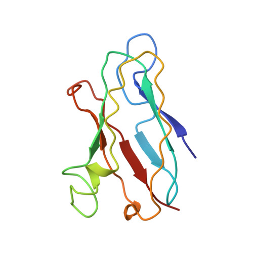
Solution structure of reduced plastocyanin from the blue-green alga Anabaena variabilis.
Badsberg, U., Jorgensen, A.M., Gesmar, H., Led, J.J., Hammerstad, J.M., Jespersen, L.L., Ulstrup, J.(1996) Biochemistry 35: 7021-7031
- PubMed: 8679527
- DOI: https://doi.org/10.1021/bi960621y
- Primary Citation of Related Structures:
1NIN - PubMed Abstract:
The three-dimensional solution structure of plastocyanin from Anabaena variabilis (A.v.PCu) has been determined by nuclear magnetic resonance spectroscopy. Sixty structures were calculated by distance geometry from 1141 distance restraints and 46 dihedral angle restraints. The distance geometry structures were optimized by simulated annealing and restrained energy minimization. The average rms deviation from the mean structure for the 20 structures with the lowest total energy is 1.25 A for the backbone atoms and 1.75 A for all heavy atoms. Overall, the global tertiary fold of A.v.PCu resembles those of other plastocyanins which have been structurally characterized by X-ray diffraction and NMR methods. This holds even though A.v.PCu is longer than any other known plastocyanins, contains far less invariant amino acid residues, and has an overall charge that differs considerably from those of other plastocyanins (+1 vs -9 +/- 1 at pH > or = 7). The most striking feature of the A.v. PCu structure is the absence of the beta-turn, formed at the remote site by residues (58)-(61) in most higher plant plastocyanins. The displacement caused by the absence of this turn is compensated for by an extension of the small helix [from Ala53(51) to Ser60(58) in A.v.PCu] found in other plastocyanins. Moreover, the extra residues of A.v.PCu from Pro77 to Asp79 form an appended loop. These two features allow A.v.PCu to retain almost the same global fold as observed in other plastocyanins. From a comparison with the structures of other plastocyanins it is concluded that the lack of negatively charged residues at the remote site, rather than the specific structure of A.v.PCu, is the main reason for the failure of the remote site of this plastocyanin to function as a significant electron transfer site.
Organizational Affiliation:
Department of Chemistry, University of Copenhagen, H. C. Orsted Institute, Denmark.