1LAU

URACIL-DNA GLYCOSYLASE


Experimental Data Snapshot

  • Method: X-RAY DIFFRACTION
  • Resolution: 1.80 Å
  • R-Value Free: 0.225 
  • R-Value Work: 0.174 
  • R-Value Observed: 0.174 

wwPDB Validation   3D Report Full Report


This is version 1.3 of the entry. See complete history


Literature

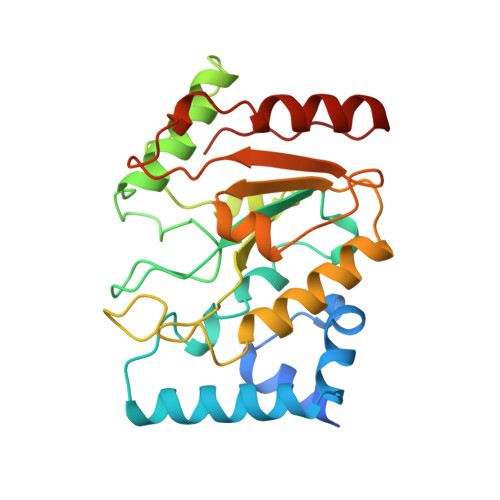
The structural basis of specific base-excision repair by uracil-DNA glycosylase.

Savva, R.McAuley-Hecht, K.Brown, T.Pearl, L.

(1995) Nature 373: 487-493

  • DOI: https://doi.org/10.1038/373487a0
  • Primary Citation of Related Structures:  
    1LAU, 1UDG, 1UDH

  • PubMed Abstract: 

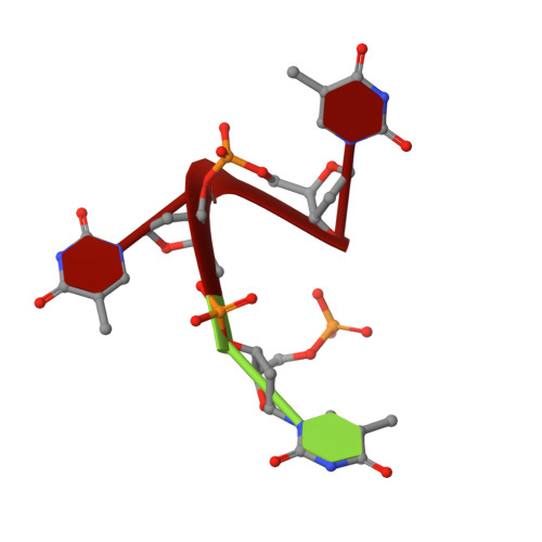
    The 1.75-A crystal structure of the uracil-DNA glycosylase from herpes simplex virus type-1 reveals a new fold, distantly related to dinucleotide-binding proteins. Complexes with a trideoxynucleotide, and with uracil, define the DNA-binding site and allow a detailed understanding of the exquisitely specific recognition of uracil in DNA. The overall structure suggests binding models for elongated single- and double-stranded DNA substrates. Conserved residues close to the uracil-binding site suggest a catalytic mechanism for hydrolytic base excision.


  • Organizational Affiliation

    Department of Biochemistry and Molecular Biology, University College London, UK.


Macromolecules

Find similar proteins by:  (by identity cutoff)  |  3D Structure
Entity ID: 2
MoleculeChains Sequence LengthOrganismDetailsImage
PROTEIN (URACIL-DNA GLYCOSYLASE (E.C.3.2.2.-))B [auth E]244Human alphaherpesvirus 1Mutation(s): 0 
UniProt
Find proteins for P10186 (Human herpesvirus 1 (strain 17))
Explore P10186 
Go to UniProtKB:  P10186
Entity Groups  
Sequence Clusters30% Identity50% Identity70% Identity90% Identity95% Identity100% Identity
UniProt GroupP10186
Sequence Annotations
Expand
  • Reference Sequence

Find similar nucleic acids by:  Sequence   |   3D Structure  

Entity ID: 1
MoleculeChains LengthOrganismImage
DNA (5'-D(*TP*TP*T)-3')A [auth D]3N/A
Sequence Annotations
Expand
  • Reference Sequence
Experimental Data & Validation

Experimental Data

  • Method: X-RAY DIFFRACTION
  • Resolution: 1.80 Å
  • R-Value Free: 0.225 
  • R-Value Work: 0.174 
  • R-Value Observed: 0.174 
  • Space Group: P 21 21 21
Unit Cell:
Length ( Å )Angle ( ˚ )
a = 46.66α = 90
b = 61.83β = 90
c = 86.67γ = 90
Software Package:
Software NamePurpose
X-PLORrefinement
MOSFLMdata reduction

Structure Validation

View Full Validation Report


Entry History 

Deposition Data

Revision History  (Full details and data files)

  • Version 1.0: 1996-06-10
    Type: Initial release
  • Version 1.1: 2008-05-22
    Changes: Version format compliance
  • Version 1.2: 2011-07-13
    Changes: Version format compliance
  • Version 1.3: 2024-02-14
    Changes: Data collection, Database references