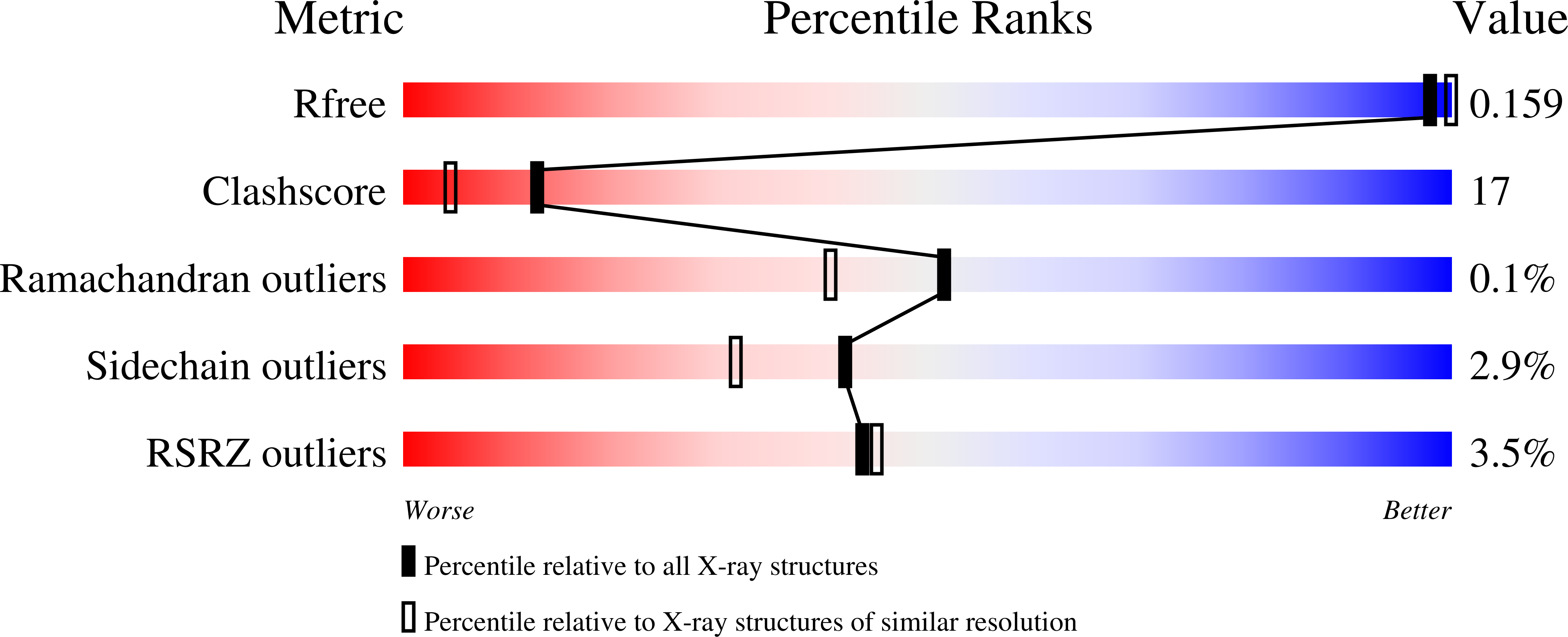
1JPR

Mn substituted Ribonucleotide reductase R2 from E. coli oxidized by nitric oxide


Experimental Data Snapshot

  • Method: X-RAY DIFFRACTION
  • Resolution: 1.88 Å
  • R-Value Free: 0.213 
  • R-Value Work: 0.158 
  • R-Value Observed: 0.160 

wwPDB Validation   3D Report Full Report


This is version 1.4 of the entry. See complete history


Literature

Crystal structures of oxidized dinuclear manganese centres in Mn-substituted class I ribonucleotide reductase from Escherichia coli: carboxylate shifts with implications for O2 activation and radical generation.

Hogbom, M.Andersson, M.E.Nordlund, P.

(2001) J Biol Inorg Chem 6: 315-323

  • DOI: https://doi.org/10.1007/s007750000205
  • Primary Citation of Related Structures:  
    1JPR, 1JQC

  • PubMed Abstract: 

    The di-iron carboxylate proteins constitute a diverse class of non-heme iron enzymes performing a multitude of redox reactions. These reactions usually involve high-valent Fe-oxo species and are thought to be controlled by carboxylate shifts. Owing to their short lifetime, the intermediate structures have so far escaped structural characterization by X-ray crystallography. In an attempt to map the carboxylate conformations available to the protein during different redox states and different ligand environments, we have studied metal-substituted forms of the R2 protein of ribonucleotide reductase from Escherichia coli. In the present work we have solved the crystal structures of Mn-substituted R2 oxidized in two different ways. Oxidation was performed using either nitric oxide or a combination of hydrogen peroxide and hydroxylamine. The two structures are virtually identical, indicating that the oxidation states are the same, most likely a mixed-valent MnII-MnIII centre. One of the carboxylate ligands (D84) adopts a new, so far unseen, conformation, which could participate in the mechanism for radical generation in R2. E238 adopts a bridging-chelating conformation proposed to be important for proper O2 activation but not previously observed in the wild-type enzyme. Probable catalase activity was also observed during the oxidation with H2O2, indicating mechanistic similarities to the di-Mn catalases.


  • Organizational Affiliation

    Department of Biochemistry and Biophysics, Arrhenius Laboratories for Natural Sciences, Stockholm University, 106 91 Stockholm, Sweden.


Macromolecules
Find similar proteins by:  (by identity cutoff)  |  3D Structure
Entity ID: 1
MoleculeChains Sequence LengthOrganismDetailsImage
Protein R2 of Ribonucleotide reductase
A, B
375Escherichia coliMutation(s): 0 
EC: 1.17.4.1
UniProt
Find proteins for P69924 (Escherichia coli (strain K12))
Explore P69924 
Go to UniProtKB:  P69924
Entity Groups  
Sequence Clusters30% Identity50% Identity70% Identity90% Identity95% Identity100% Identity
UniProt GroupP69924
Sequence Annotations
Expand
  • Reference Sequence
Small Molecules
Ligands 2 Unique
IDChains Name / Formula / InChI Key2D Diagram3D Interactions
HG
Query on HG

Download Ideal Coordinates CCD File 
E [auth A]
F [auth A]
G [auth A]
H [auth A]
K [auth B]
E [auth A],
F [auth A],
G [auth A],
H [auth A],
K [auth B],
L [auth B],
M [auth B],
N [auth B],
O [auth B],
P [auth B],
Q [auth B],
R [auth B],
S [auth B],
T [auth B]
MERCURY (II) ION
Hg
BQPIGGFYSBELGY-UHFFFAOYSA-N
MN
Query on MN

Download Ideal Coordinates CCD File 
C [auth A],
D [auth A],
I [auth B],
J [auth B]
MANGANESE (II) ION
Mn
WAEMQWOKJMHJLA-UHFFFAOYSA-N
Experimental Data & Validation

Experimental Data

  • Method: X-RAY DIFFRACTION
  • Resolution: 1.88 Å
  • R-Value Free: 0.213 
  • R-Value Work: 0.158 
  • R-Value Observed: 0.160 
  • Space Group: P 21 21 21
Unit Cell:
Length ( Å )Angle ( ˚ )
a = 73.826α = 90
b = 84.683β = 90
c = 114.333γ = 90
Software Package:
Software NamePurpose
CNSrefinement
DENZOdata reduction
SCALEPACKdata scaling
CNSphasing

Structure Validation

View Full Validation Report


Entry History 

Deposition Data

Revision History  (Full details and data files)

  • Version 1.0: 2001-08-15
    Type: Initial release
  • Version 1.1: 2008-04-27
    Changes: Version format compliance
  • Version 1.2: 2011-07-13
    Changes: Version format compliance
  • Version 1.3: 2024-03-13
    Changes: Data collection, Database references, Derived calculations
  • Version 1.4: 2024-04-03
    Changes: Refinement description