Insights into the structure, solvation, and mechanism of ArsC arsenate reductase, a novel arsenic detoxification enzyme.
Martin, P., DeMel, S., Shi, J., Gladysheva, T., Gatti, D.L., Rosen, B.P., Edwards, B.F.(2001) Structure 9: 1071-1081
- PubMed: 11709171
- DOI: https://doi.org/10.1016/s0969-2126(01)00672-4
- Primary Citation of Related Structures:
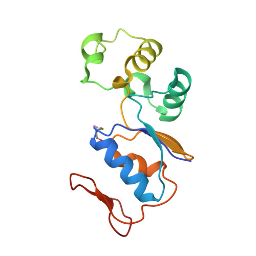
1I9D, 1J9B, 1JZW - PubMed Abstract:
In Escherichia coli bearing the plasmid R773, resistance to arsenite, arsenate, antimonite, and tellurite is conferred by the arsRDABC plasmid operon that codes for an ATP-dependent anion pump. The product of the arsC gene, arsenate reductase (ArsC), is required to efficiently catalyze the reduction of arsenate to arsenite prior to extrusion. Here, we report the first X-ray crystal structures of ArsC at 1.65 A and of ArsC complexed with arsenate and arsenite at 1.26 A resolution. The overall fold is unique. The native structure shows sulfate and sulfite ions binding in the active site as analogs of arsenate and arsenite. The covalent adduct of arsenate with Cys-12 in the active site of ArsC, which was analyzed in a difference map, shows tetrahedral geometry with a sulfur-arsenic distance of 2.18 A. However, the corresponding adduct with arsenite binds as a hitherto unseen thiarsahydroxy adduct. Finally, the number of bound waters (385) in this highly ordered crystal structure approaches twice the number expected at this resolution for a structure of 138 ordered residues. Structural information from the adduct of ArsC with its substrate (arsenate) and with its product (arsenite) together with functional information from mutational and biochemical studies on ArsC suggest a plausible mechanism for the reaction. The exceptionally well-defined water structure indicates that this crystal system has precise long-range order within the crystal and that the upper limit for the number of bound waters in crystal structures is underestimated by the structures in the Protein Data Bank.
Organizational Affiliation:
Wayne State University School of Medicine, Department of Biochemistry and Molecular Biology, 540 E. Canfield, Detroit, MI 48201, USA.