1J77

Crystal Structure of Gram-negative Bacterial Heme Oxygenase Complexed with Heme


Experimental Data Snapshot

  • Method: X-RAY DIFFRACTION
  • Resolution: 1.50 Å
  • R-Value Free: 0.260 
  • R-Value Work: 0.227 
  • R-Value Observed: 0.229 

wwPDB Validation   3D Report Full Report


Ligand Structure Quality Assessment 


This is version 1.3 of the entry. See complete history


Literature

Crystal structure of heme oxygenase from the gram-negative pathogen Neisseria meningitidis and a comparison with mammalian heme oxygenase-1.

Schuller, D.J.Zhu, W.Stojiljkovic, I.Wilks, A.Poulos, T.L.

(2001) Biochemistry 40: 11552-11558

  • DOI: https://doi.org/10.1021/bi0110239
  • Primary Citation of Related Structures:  
    1J77

  • PubMed Abstract: 

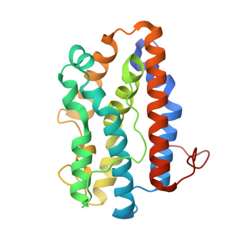
    We report the crystal structure of heme oxygenase from the pathogenic bacterium Neisseria meningitidis at 1.5 A and compare and contrast it with known structures of heme oxygenase-1 from mammalian sources. Both the bacterial and mammalian enzymes share the same overall fold, with a histidine contributing a ligand to the proximal side of the heme iron and a kinked alpha-helix defining the distal pocket. The distal helix differs noticeably in both sequence and conformation, and the distal pocket of the Neisseria enzyme is substantially smaller than in the mammalian enzyme. Key glycine residues provide the flexibility for the helical kink, allow close contact of the helix backbone with the heme, and may interact directly with heme ligands.


  • Organizational Affiliation

    Department of Molecular Biology & Biochemistry, Department of Physiology & Biophysics, and Program in Structural Biology, University of California, Irvine, California 92697-3900, USA.


Macromolecules
Find similar proteins by:  (by identity cutoff)  |  3D Structure
Entity ID: 1
MoleculeChains Sequence LengthOrganismDetailsImage
HemO209Neisseria meningitidisMutation(s): 0 
Gene Names: hemO
EC: 1.14.99.3
UniProt
Find proteins for Q9RGD9 (Neisseria meningitidis)
Explore Q9RGD9 
Go to UniProtKB:  Q9RGD9
Entity Groups  
Sequence Clusters30% Identity50% Identity70% Identity90% Identity95% Identity100% Identity
UniProt GroupQ9RGD9
Sequence Annotations
Expand
  • Reference Sequence
Small Molecules
Ligands 1 Unique
IDChains Name / Formula / InChI Key2D Diagram3D Interactions
HEM
Query on HEM

Download Ideal Coordinates CCD File 
B [auth A]PROTOPORPHYRIN IX CONTAINING FE
C34 H32 Fe N4 O4
KABFMIBPWCXCRK-RGGAHWMASA-L
Experimental Data & Validation

Experimental Data

  • Method: X-RAY DIFFRACTION
  • Resolution: 1.50 Å
  • R-Value Free: 0.260 
  • R-Value Work: 0.227 
  • R-Value Observed: 0.229 
  • Space Group: P 43 21 2
Unit Cell:
Length ( Å )Angle ( ˚ )
a = 63.167α = 90
b = 63.167β = 90
c = 100.378γ = 90
Software Package:
Software NamePurpose
SHARPphasing
REFMACrefinement
DENZOdata reduction
SCALEPACKdata scaling

Structure Validation

View Full Validation Report


Ligand Structure Quality Assessment 


Entry History 

Deposition Data

Revision History  (Full details and data files)

  • Version 1.0: 2001-05-30
    Type: Initial release
  • Version 1.1: 2008-04-27
    Changes: Version format compliance
  • Version 1.2: 2011-07-13
    Changes: Version format compliance
  • Version 1.3: 2024-02-07
    Changes: Data collection, Database references, Derived calculations