1IH0

Structure of the C-domain of Human Cardiac Troponin C in Complex with Ca2+ Sensitizer EMD 57033


Experimental Data Snapshot

  • Method: SOLUTION NMR
  • Conformers Calculated: 30 
  • Conformers Submitted: 30 
  • Selection Criteria: all calculated structures submitted 

wwPDB Validation   3D Report Full Report


This is version 1.3 of the entry. See complete history


Literature

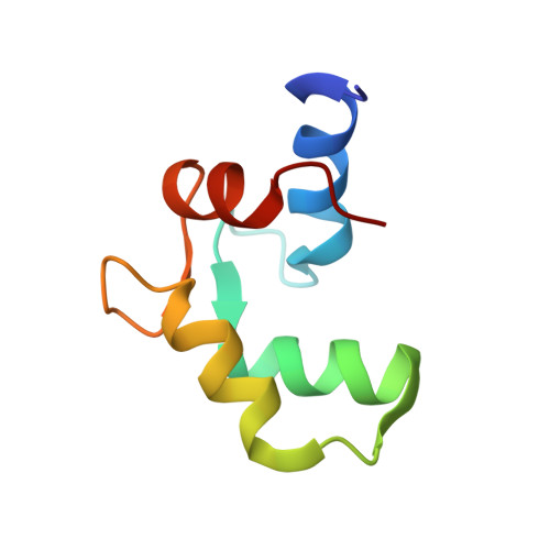
Structure of the C-domain of human cardiac troponin C in complex with the Ca2+ sensitizing drug EMD 57033.

Wang, X.Li, M.X.Spyracopoulos, L.Beier, N.Chandra, M.Solaro, R.J.Sykes, B.D.

(2001) J Biol Chem 276: 25456-25466

  • DOI: https://doi.org/10.1074/jbc.M102418200
  • Primary Citation of Related Structures:  
    1IH0

  • PubMed Abstract: 

    Ca(2+) binding to cardiac troponin C (cTnC) triggers contraction in heart muscle. In heart failure, myofilaments response to Ca(2+) are often altered and compounds that sensitize the myofilaments to Ca(2+) possess therapeutic value in this syndrome. One of the most potent and selective Ca(2+) sensitizers is the thiadiazinone derivative EMD 57033, which increases myocardial contractile function both in vivo and in vitro and interacts with cTnC in vitro. We have determined the NMR structure of the 1:1 complex between Ca(2+)-saturated C-domain of human cTnC (cCTnC) and EMD 57033. Favorable hydrophobic interactions between the drug and the protein position EMD 57033 in the hydrophobic cleft of the protein. The drug molecule is orientated such that the chiral group of EMD 57033 fits deep in the hydrophobic pocket and makes several key contacts with the protein. This stereospecific interaction explains why the (-)-enantiomer of EMD 57033 is inactive. Titrations of the cCTnC.EMD 57033 complex with two regions of cardiac troponin I (cTnI(34-71) and cTnI(128-147)) reveal that the drug does not share a common binding epitope with cTnI(128-147) but is completely displaced by cTnI(34-71). These results have important implications for elucidating the mechanism of the Ca(2+) sensitizing effect of EMD 57033 in cardiac muscle contraction.


  • Organizational Affiliation

    CIHR Group in Protein Structure and Function, Department of Biochemistry, University of Alberta, Edmonton, Alberta T6G 2H7, Canada.


Macromolecules
Find similar proteins by:  (by identity cutoff)  |  3D Structure
Entity ID: 1
MoleculeChains Sequence LengthOrganismDetailsImage
TROPONIN C, SLOW SKELETAL AND CARDIAC MUSCLES71Homo sapiensMutation(s): 0 
Gene Names: cTnC
UniProt & NIH Common Fund Data Resources
Find proteins for P63316 (Homo sapiens)
Explore P63316 
Go to UniProtKB:  P63316
PHAROS:  P63316
GTEx:  ENSG00000114854 
Entity Groups  
Sequence Clusters30% Identity50% Identity70% Identity90% Identity95% Identity100% Identity
UniProt GroupP63316
Sequence Annotations
Expand
  • Reference Sequence
Small Molecules
Ligands 2 Unique
IDChains Name / Formula / InChI Key2D Diagram3D Interactions
EMD
Query on EMD

Download Ideal Coordinates CCD File 
D [auth A]5-[1-(3,4-DIMETHOXY-BENZOYL)-1,2,3,4-TETRAHYDRO-QUINOLIN-6-YL]-6-METHYL-3,6-DIHYDRO-[1,3,4]THIADIAZIN-2-ONE
C22 H23 N3 O4 S
IZLRMTJLQCLMKF-CYBMUJFWSA-N
CA
Query on CA

Download Ideal Coordinates CCD File 
B [auth A],
C [auth A]
CALCIUM ION
Ca
BHPQYMZQTOCNFJ-UHFFFAOYSA-N
Binding Affinity Annotations 
IDSourceBinding Affinity
EMD PDBBind:  1IH0 Kd: 1.00e+4 (nM) from 1 assay(s)
Experimental Data & Validation

Experimental Data

  • Method: SOLUTION NMR
  • Conformers Calculated: 30 
  • Conformers Submitted: 30 
  • Selection Criteria: all calculated structures submitted 

Structure Validation

View Full Validation Report


Entry History 

Deposition Data

Revision History  (Full details and data files)

  • Version 1.0: 2001-10-10
    Type: Initial release
  • Version 1.1: 2008-04-27
    Changes: Version format compliance
  • Version 1.2: 2011-07-13
    Changes: Version format compliance
  • Version 1.3: 2022-02-23
    Changes: Database references, Derived calculations