1HV6

CRYSTAL STRUCTURE OF ALGINATE LYASE A1-III COMPLEXED WITH TRISACCHARIDE PRODUCT.


Experimental Data Snapshot

  • Method: X-RAY DIFFRACTION
  • Resolution: 2.00 Å
  • R-Value Free: 0.197 
  • R-Value Work: 0.158 
  • R-Value Observed: 0.158 

wwPDB Validation   3D Report Full Report


This is version 2.1 of the entry. See complete history


Literature

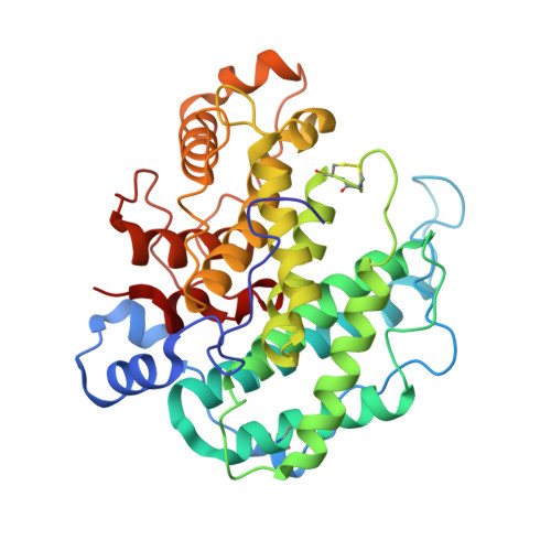
Crystal structure of alginate lyase A1-III complexed with trisaccharide product at 2.0 A resolution.

Yoon, H.J.Hashimoto, W.Miyake, O.Murata, K.Mikami, B.

(2001) J Mol Biol 307: 9-16

  • DOI: https://doi.org/10.1006/jmbi.2000.4509
  • Primary Citation of Related Structures:  
    1HV6

  • PubMed Abstract: 

    The structure of A1-III from a Sphingomonas species A1 complexed with a trisaccharide product (4-deoxy-l-erythro-hex-4-enepyranosyluronate-mannuronate-mannuronic acid) was determined by X-ray crystallography at 2.0 A with an R-factor of 0.16. The final model of the complex form comprising 351 amino acid residues, 245 water molecules, one sulfate ion and one trisaccharide product exhibited a C(alpha) r.m.s.d. value of 0.154 A with the reported apo form of the enzyme. The trisaccharide was bound in the active cleft at subsites -3 approximately -1 from the non-reducing end by forming several hydrogen bonds and van der Waals interactions with protein atoms. The catalytic residue was estimated to be Tyr246, which existed between subsites -1 and +1 based on a mannuronic acid model oriented at subsite +1.


  • Organizational Affiliation

    Research Institute for Food Science, Kyoto University, Uji Kyoto 611-0011, Japan.


Macromolecules
Find similar proteins by:  (by identity cutoff)  |  3D Structure
Entity ID: 1
MoleculeChains Sequence LengthOrganismDetailsImage
ALGINATE LYASE351Sphingomonas sp.Mutation(s): 0 
EC: 4.2.2.3
UniProt
Find proteins for Q9KWU1 (Sphingomonas sp)
Explore Q9KWU1 
Go to UniProtKB:  Q9KWU1
Entity Groups  
Sequence Clusters30% Identity50% Identity70% Identity90% Identity95% Identity100% Identity
UniProt GroupQ9KWU1
Sequence Annotations
Expand
  • Reference Sequence
Oligosaccharides

Help

Entity ID: 2
MoleculeChains Length2D Diagram Glycosylation3D Interactions
4-deoxy-alpha-L-erythro-hex-4-enopyranuronic acid-(1-4)-alpha-D-mannopyranuronic acid-(1-4)-alpha-D-glucopyranuronic acid
B
3N/A
Glycosylation Resources
GlyTouCan:  G62159RG
GlyCosmos:  G62159RG
Small Molecules
Ligands 1 Unique
IDChains Name / Formula / InChI Key2D Diagram3D Interactions
SO4
Query on SO4

Download Ideal Coordinates CCD File 
C [auth A]SULFATE ION
O4 S
QAOWNCQODCNURD-UHFFFAOYSA-L
Experimental Data & Validation

Experimental Data

  • Method: X-RAY DIFFRACTION
  • Resolution: 2.00 Å
  • R-Value Free: 0.197 
  • R-Value Work: 0.158 
  • R-Value Observed: 0.158 
  • Space Group: C 1 2 1
Unit Cell:
Length ( Å )Angle ( ˚ )
a = 48.765α = 90
b = 92.92β = 104.11
c = 82.22γ = 90
Software Package:
Software NamePurpose
SADIE/SAINTdata collection
SAINTdata reduction
CNSrefinement
SADIEdata reduction
SAINTdata scaling

Structure Validation

View Full Validation Report


Entry History 

Deposition Data

Revision History  (Full details and data files)

  • Version 1.0: 2001-05-02
    Type: Initial release
  • Version 1.1: 2008-04-27
    Changes: Version format compliance
  • Version 1.2: 2011-07-13
    Changes: Non-polymer description, Version format compliance
  • Version 2.0: 2020-07-29
    Type: Remediation
    Reason: Carbohydrate remediation
    Changes: Advisory, Atomic model, Data collection, Database references, Derived calculations, Structure summary
  • Version 2.1: 2023-10-25
    Changes: Data collection, Database references, Refinement description, Structure summary