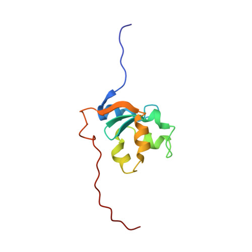
The Solution Structure of Bovine Ferricytochrome B5 Determined Using Heteronuclear NMR Methods.
Muskett, F.W., Kelly, G.P., Whitford, D.(1996) J Mol Biol 258: 172
- PubMed: 8613986
- DOI: https://doi.org/10.1006/jmbi.1996.0241
- Primary Citation of Related Structures:
1HKO - PubMed Abstract:
The solution structure of a recombinant form of cytochrome b5 containing 104 amino acid residues has been determined using three-dimensional NMR spectroscopy. Using protein enriched in 15N the majority of the polypeptide backbone resonances have been assigned to reveal numerous chemical shift differences to those reported previously for smaller fragments of cytochrome b5. By using 3D NMR methods the extensive spectral overlap of resonance cross-peaks in 2D NMR spectra could be satisfactorily resolved. The large number of sequence-specific assignments made for this form of the protein allowed the identification of over 1130 NOEs, giving an average of 14 NOEs per assigned residue, and 52 dihedral angles (phi). This data was used in an ab initio simulated annealing protocol to determine the solution structure for bovine microsomal cytochrome b5. A series of 50 structures was generated using distance restraints derived from the magnitude of the NOE and torsional angles based on the measured JHN-HA coupling constants. From an initial round of simulated annealing a family of 36 structures was selected on the basis of good covalent geometry and minimal restraint violations. A single cycle of simulated annealing refinement produced 36 converged structures that exhibited an average r.m.s.d. of 0.73 A for the backbone atoms. The determination of the solution structure of cytochrome b5 is the first using NMR methods for any form of this protein. It is also the only cytochrome whose structure has been determined in the oxidised or paramagnetic state. The results show that despite significant line broadening and pseudocontact shifts for resonances lying close to the paramagnetic haem centre, and despite extensive spectral overlap that prevents complete resonance assignment, the topology of the polypeptide backbone can be derived. The conformation for cytochrome b5 determined in this study reveals several small, but significant, differences in structure to that determined previously by crystallography for a smaller fragment of this protein. For example, NMR data do not support a short beta strand as the first element of secondary structure at the N terminus nor is it likely that a beta-bulge structure forms between residues 75 to 79. The data obtained in this study are more consistent with a turn in this region of the protein linking helices 5 and 6 and leads to cytochrome b5 containing only three clearly defined beta strands. Four of the six helices together with the antiparallel beta strands make up a haem binding pocket in which the solvent-accessible area of the protoporphyrin IX centre remains very similar to that found in the crystal structure. The remaining helices and the beta strands form a second structural domain on which the four helix bundle that surrounds the haem is based. THe derivation of the solution structure of cytochrome b5 will allow a greater understanding of the functional properties of cytochrome b5 including its role in biological electron transfer and molecular recognition together with insight into haem protein folding and stability.
Organizational Affiliation:
Department of Biochemistry, Queen Mary and Westfield College, London.