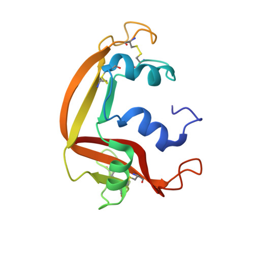
1H1H

Crystal Structure of Eosinophil Cationic Protein in Complex with 2',5'-ADP at 2.0 A resolution Reveals the Details of the Ribonucleolytic Active site


Experimental Data Snapshot

  • Method: X-RAY DIFFRACTION
  • Resolution: 2.00 Å
  • R-Value Free: 0.210 
  • R-Value Work: 0.205 
  • R-Value Observed: 0.205 

wwPDB Validation   3D Report Full Report


This is version 1.4 of the entry. See complete history


Literature

The Crystal Structure of Eosinophil Cationic Protein in Complex with 2'5'-Adp at 2.0 A Resolution Reveals the Details of the Ribonucleolytic Active Site

Mohan, C.G.Boix, E.Evans, H.R.Nikolovski, Z.Nogues, M.V.Cuchillo, C.M.Acharya, K.R.

(2002) Biochemistry 41: 12100

  • DOI: https://doi.org/10.1021/bi0264521
  • Primary Citation of Related Structures:  
    1H1H

  • PubMed Abstract: 

    Eosinophil cationic protein (ECP) is a component of the eosinophil granule matrix. It shows marked toxicity against helminth parasites, bacteria single-stranded RNA viruses, and host epithelial cells. Secretion of human ECP is related to eosinophil-associated allergic, asthmatic, and inflammatory diseases. ECP belongs to the pancreatic ribonuclease superfamily of proteins, and the crystal structure of ECP in the unliganded form (determined previously) exhibited a conserved RNase A fold [Boix, E., et al. (1999) Biochemistry 38, 16794-16801]. We have now determined a high-resolution (2.0 A) crystal structure of ECP in complex with adenosine 2',5'-diphosphate (2',5'-ADP) which has revealed the details of the ribonucleolytic active site. Residues Gln-14, His-15, and Lys-38 make hydrogen bond interactions with the phosphate at the P(1) site, while His-128 interacts with the purine ring at the B(2) site. A new phosphate binding site, P(-)(1), has been identified which involves Arg-34. This study is the first detailed structural analysis of the nucleotide recognition site in ECP and provides a starting point for the understanding of its substrate specificity and low catalytic efficiency compared with that of the eosinophil-derived neurotoxin (EDN), a close homologue.


  • Organizational Affiliation

    Department of Biology and Biochemistry, University of Bath, Claverton Down, Bath BA2 7AY, U.K.


Macromolecules
Find similar proteins by:  (by identity cutoff)  |  3D Structure
Entity ID: 1
MoleculeChains Sequence LengthOrganismDetailsImage
EOSINOPHIL CATIONIC PROTEIN134Homo sapiensMutation(s): 0 
EC: 3.1.27
Membrane Entity: Yes 
UniProt & NIH Common Fund Data Resources
Find proteins for P12724 (Homo sapiens)
Explore P12724 
Go to UniProtKB:  P12724
PHAROS:  P12724
GTEx:  ENSG00000169397 
Entity Groups  
Sequence Clusters30% Identity50% Identity70% Identity90% Identity95% Identity100% Identity
UniProt GroupP12724
Sequence Annotations
Expand
  • Reference Sequence
Small Molecules
Ligands 1 Unique
IDChains Name / Formula / InChI Key2D Diagram3D Interactions
A2P
Query on A2P

Download Ideal Coordinates CCD File 
B [auth A]ADENOSINE-2'-5'-DIPHOSPHATE
C10 H15 N5 O10 P2
AEOBEOJCBAYXBA-KQYNXXCUSA-N
Binding Affinity Annotations 
IDSourceBinding Affinity
A2P PDBBind:  1H1H Ki: 6000 (nM) from 1 assay(s)
Binding MOAD:  1H1H Ki: 6500 (nM) from 1 assay(s)
Experimental Data & Validation

Experimental Data

  • Method: X-RAY DIFFRACTION
  • Resolution: 2.00 Å
  • R-Value Free: 0.210 
  • R-Value Work: 0.205 
  • R-Value Observed: 0.205 
  • Space Group: P 63
Unit Cell:
Length ( Å )Angle ( ˚ )
a = 100.7α = 90
b = 100.7β = 90
c = 31.7γ = 120
Software Package:
Software NamePurpose
CNSrefinement
DENZOdata reduction
SCALEPACKdata scaling
AMoREphasing

Structure Validation

View Full Validation Report


Entry History 

Deposition Data

Revision History  (Full details and data files)

  • Version 1.0: 2002-10-03
    Type: Initial release
  • Version 1.1: 2011-05-08
    Changes: Version format compliance
  • Version 1.2: 2011-07-13
    Changes: Version format compliance
  • Version 1.3: 2019-10-16
    Changes: Data collection
  • Version 1.4: 2023-12-13
    Changes: Data collection, Database references, Refinement description