Nickel-guanine interactions in DNA: crystal structure of nickel-d[CGTGTACACG]2.
Abrescia, N.A., Huynh-Dinh, T., Subirana, J.A.(2002) J Biol Inorg Chem 7: 195-199
- PubMed: 11862555
- DOI: https://doi.org/10.1007/s007750100286
- Primary Citation of Related Structures:
1G3V - PubMed Abstract:
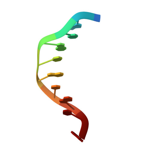
The aim of this study was to clarify whether Ni2+ ions could bind to guanine bases in a standard B-DNA duplex and eventually induce a B-->Z transition. We have determined by X-ray crystallography at 3.1 A resolution the structure of the alternating deoxynucleotide d(CGTGTACACG), which contains both internal and terminal guanines. The duplex is in the B form. It is shown that nickel ions bind selectively to the N7 atom of guanine 10, which is in an extra-helical position, and guanine 2, which is in the terminal position of the duplex. It does not bind to guanine 4, which lies within a standard B-DNA tract. This simple but unambiguous result proves that nickel ions select between different guanines via steric accessibility. Guanine-Ni2+-guanine bridges among symmetry-related duplexes have also been found. These bridges may explain why Ni2+ ions may act either as a precipitant or a renaturing agent for DNA under certain conditions. The biochemical interaction of nickel with DNA can thus be related to its capacity to specifically bind to B-DNA regions with exposed guanines. Also, from the structural point of view, we have found a terminal cytosine, which forms a C.G:C reverse-Hoogsteen triple structure with a base pair of a neighbor duplex. This type of triplet is seldom found and is here described for the first time for a DNA structure.
Organizational Affiliation:
Departament d'Enginyeria Química, Universitat Politècnica de Catalunya, Diagonal 647, 08028 Barcelona, Spain.