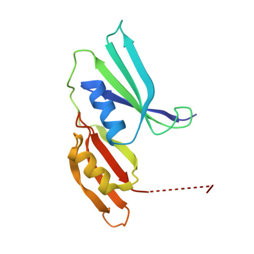
Determination of the structure of Escherichia coli glyoxalase I suggests a structural basis for differential metal activation.
He, M.M., Clugston, S.L., Honek, J.F., Matthews, B.W.(2000) Biochemistry 39: 8719-8727
- PubMed: 10913283
- DOI: https://doi.org/10.1021/bi000856g
- Primary Citation of Related Structures:
1F9Z, 1FA5, 1FA6, 1FA7, 1FA8 - PubMed Abstract:
The metalloenzyme glyoxalase I (GlxI) converts the nonenzymatically produced hemimercaptal of cytotoxic methylglyoxal and glutathione to nontoxic S-D-lactoylglutathione. Human GlxI, for which the structure is known, is active in the presence of Zn(2+). Unexpectedly, the Escherichia coli enzyme is inactive in the presence of Zn(2+) and is maximally active with Ni(2+). To understand this difference in metal activation and also to obtain a representative of the bacterial enzymes, the structure of E. coli Ni(2+)-GlxI has been determined. Structures have also been determined for the apo enzyme as well as complexes with Co(2+), Cd(2+), and Zn(2+). It is found that each of the protein-metal complexes that is catalytically active has octahedral geometry. This includes the complexes of the E. coli enzyme with Ni(2+), Co(2+), and Cd(2+), as well as the structures reported for the human Zn(2+) enzyme. Conversely, the complex of the E. coli enzyme with Zn(2+) has trigonal bipyramidal coordination and is inactive. This mode of coordination includes four protein ligands plus a single water molecule. In contrast, the coordination in the active forms of the enzyme includes two water molecules bound to the metal ion, suggesting that this may be a key feature of the catalytic mechanism. A comparison of the human and E. coli enzymes suggests that there are differences between the active sites that might be exploited for therapeutic use.
Organizational Affiliation:
Howard Hughes Medical Institute, Institute of Molecular Biology, Department of Physics, 1229 University of Oregon, Eugene, Oregon 97403-1229, USA.