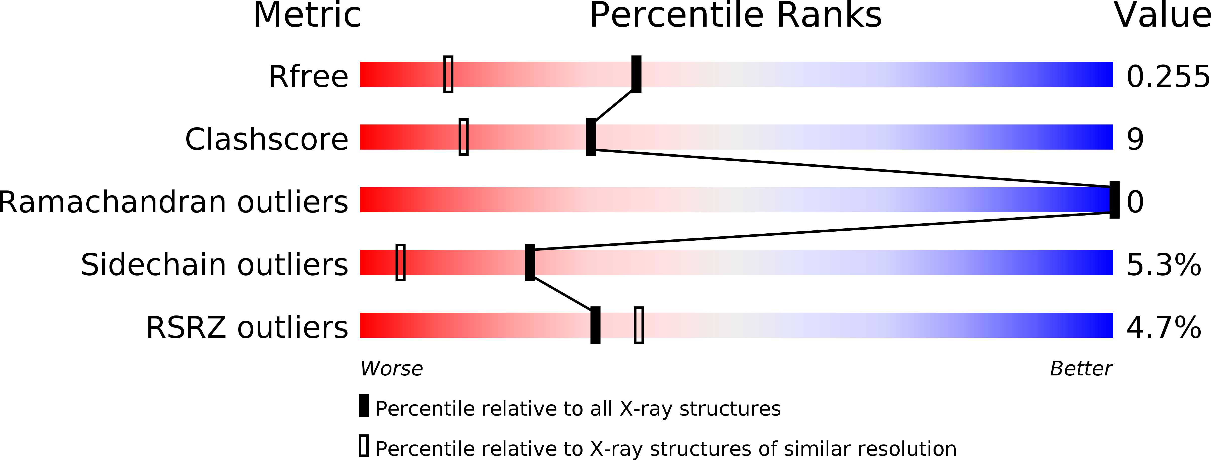
Structural basis for the inhibition of porcine pepsin by Ascaris pepsin inhibitor-3.
Ng, K.K., Petersen, J.F., Cherney, M.M., Garen, C., Zalatoris, J.J., Rao-Naik, C., Dunn, B.M., Martzen, M.R., Peanasky, R.J., James, M.N.(2000) Nat Struct Biol 7: 653-657
- PubMed: 10932249
- DOI: https://doi.org/10.1038/77950
- Primary Citation of Related Structures:
1F32, 1F34 - PubMed Abstract:
The three-dimensional structures of pepsin inhibitor-3 (PI-3) from Ascaris suum and of the complex between PI-3 and porcine pepsin at 1. 75 A and 2.45 A resolution, respectively, have revealed the mechanism of aspartic protease inhibition by this unique inhibitor. PI-3 has a new fold consisting of two domains, each comprising an antiparallel beta-sheet flanked by an alpha-helix. In the enzyme-inhibitor complex, the N-terminal beta-strand of PI-3 pairs with one strand of the 'active site flap' (residues 70-82) of pepsin, thus forming an eight-stranded beta-sheet that spans the two proteins. PI-3 has a novel mode of inhibition, using its N-terminal residues to occupy and therefore block the first three binding pockets in pepsin for substrate residues C-terminal to the scissile bond (S1'-S3'). The molecular structure of the pepsin-PI-3 complex suggests new avenues for the rational design of proteinaceous aspartic proteinase inhibitors.
Organizational Affiliation:
MRC of Canada, Group in Protein Structure and Function, Department of Biochemistry, University of Alberta, Edmonton, Alberta T6G 2H7, Canada.


