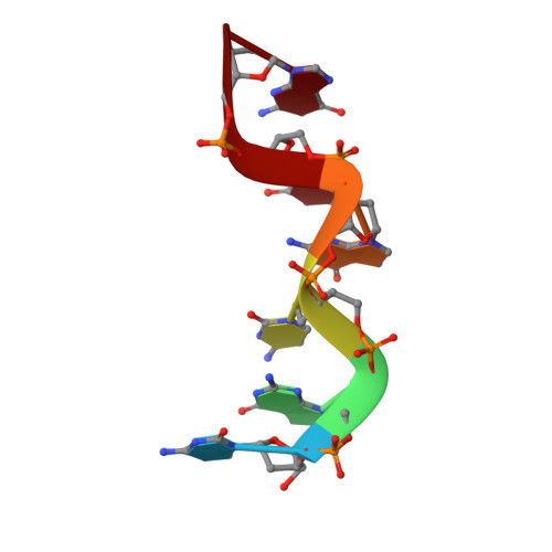
Covalent modification of guanine bases in double-stranded DNA. The 1.2-A Z-DNA structure of d(CGCGCG) in the presence of CuCl2.
Kagawa, T.F., Geierstanger, B.H., Wang, A.H., Ho, P.S.(1991) J Biol Chem 266: 20175-20184
- PubMed: 1939078
- DOI: https://doi.org/10.2210/pdb1d39/pdb
- Primary Citation of Related Structures:
1D39 - PubMed Abstract:
We have solved the single crystal structure to 1.2-A resolution of the Z-DNA sequence d(CGCGCG) soaked with copper(II) chloride. This structure allows us to elucidate the structural properties of copper in a model that mimics a physiologically relevant environment. A copper(II) cation was observed to form a covalent coordinate bond to N-7 of each guanine base along the hexamer duplex. The occurrence of copper bound at each site was dependent on the exposure of the bases and the packing of the hexamers in the crystal. The copper at the highest occupied site was observed to form a regular octahedral complex, with four water ligands in the equatorial plane and a fifth water along with N-7 of the purine base at the axial positions. All other copper complexes appear to be variations of this structure. By using the octahedral complex as the prototype for copper(II) binding to guanine bases in the Z-DNA crystal, model structures were built showing that duplex B-DNA can accommodate octahedral copper(II) complexes at the guanine bases as well as copper complexes bridged at adjacent guanine residues by a reactive dioxygen species. The increased susceptibility to oxidative DNA cleavage induced by copper(II) ions in solution of the bases located 5' to one or more adjacent guanine residues can thus be explained in terms of the cation and DNA structures described by these models.
Organizational Affiliation:
Department of Biochemistry and Biophysics, Oregon State University, Corvallis 97331-6503.