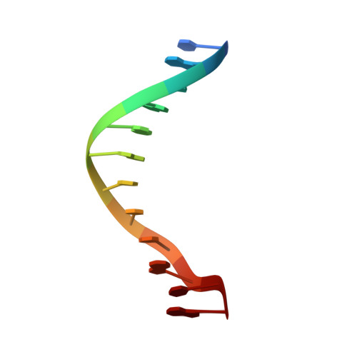
The structure of DAPI bound to DNA.
Larsen, T.A., Goodsell, D.S., Cascio, D., Grzeskowiak, K., Dickerson, R.E.(1989) J Biomol Struct Dyn 7: 477-491
- PubMed: 2627296
- DOI: https://doi.org/10.1080/07391102.1989.10508505
- Primary Citation of Related Structures:
1D30 - PubMed Abstract:
The structure of the DNA fluorochrome 4'-6-diamidine-2-phenyl indole (DAPI) bound to the synthetic B-DNA oligonucleotide C-G-C-G-A-A-T-T-C-G-C-G has been solved by single crystal x-ray diffraction methods, at a resolution of 2.4 A. The structure is nearly isomorphous with that of the native DNA molecule alone. With one DAPI and 25 waters per DNA double helix, the residual error is 21.5% for the 2428 reflections above the 2-sigma level. DAPI inserts itself edgewise into the narrow minor groove, displacing the ordered spine of hydration. DAPI and a single water molecule together span the four AT base pairs at the center of the duplex. The indole nitrogen forms a bifurcated hydrogen bond with the thymine O2 atoms of the two central base pairs, as with netropsin and Hoechst 33258. The preference of all three of these drugs for AT regions of B-DNA is a consequence of three factors: (1) The intrinsically narrower minor groove in AT regions than in GC regions of B-DNA, leading to a snug fit of the flat aromatic drug rings between the walls of the groove. (2) The more negative electrostatic potential within the minor groove in AT regions, attributable in part to the absence of electropositive-NH2 groups along the floor of the groove, and (3) The steric advantage of the absence of those same guanine-NH2 groups, thus permitting the drug molecule to sink deeper into the groove. Groove width and electrostatic factors are regional, and define the relative receptiveness of a section of DNA since they operate over several contiguous base pairs. The steric factor is local, varying from one base pair to the next, and hence is the means of fine-tuning sequence specificity.
Organizational Affiliation:
Molecular Biology Institute, University of California, Los Angeles 90024-1570.