Binding of the antitumor drug nogalamycin and its derivatives to DNA: structural comparison.
Gao, Y.G., Liaw, Y.C., Robinson, H., Wang, A.H.(1990) Biochemistry 29: 10307-10316
- PubMed: 2261474
- DOI: https://doi.org/10.1021/bi00497a004
- Primary Citation of Related Structures:
1D21, 1D22 - PubMed Abstract:
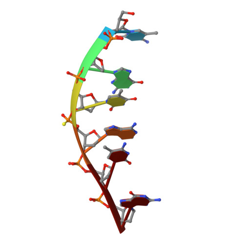
The three-dimensional molecular structures of the complexes between a novel antitumor drug nogalamycin and its derivative U-58872 with a modified DNA hexamer d[m5CGT(pS)Am5CG] have been determined at 1.7- and 1.8-A resolution, respectively, by X-ray diffraction analyses. Both structures (in space group P6(1)) have been refined with constrained refinement procedure to final R factors of 0.208 (3386 reflections) and 0.196 (2143 reflections). In both complexes, two nogalamycins bind to the DNA hexamer double helix in a 2:1 ratio with the elongated aglycon chromophore intercalated between the CpG steps at both ends of the helix. The aglycon chromophore spans across the GC Watson-Crick base pairs with its nogalose lying in the minor groove and the aminoglucose lying in the major groove of the distorted B-DNA double helix. Most of the sugars remain in the C2'-endo pucker family, except three deoxycytidine residues (terminal C1, C7, and internal C5). All nucleotides are in the anti conformation. Specific hydrogen bonds are found in the complex between the drug and guanine-cytosine bases in both grooves of the helix. One hydroxyl group of the aminoglucose donates a hydrogen bond to the N7 of guanine, while the other receives a hydrogen bond from the N4 amino group of cytosine. The orientation of these two hydrogen bonds suggests that nogalamycin prefers a GC base pair with its aglycon chromophore intercalating at the 5'-side of a guanine (between NpG), or at the 3'-side of a cytosine (between CpN) with the sugars pointing toward the GC base pair. The binding of nogalamycin to DNA requires that the base pairs in DNA open up transiently to allow the bulky sugars to go through, suggesting that nogalamycin prefers GC sequences embedded in a stretch of AT sequences.
Organizational Affiliation:
Department of Physiology and Biophysics, University of Illinois, Urbana 61801.