1D0Q

STRUCTURE OF THE ZINC-BINDING DOMAIN OF BACILLUS STEAROTHERMOPHILUS DNA PRIMASE


Experimental Data Snapshot

  • Method: X-RAY DIFFRACTION
  • Resolution: 1.71 Å
  • R-Value Free: 0.225 
  • R-Value Work: 0.204 

wwPDB Validation   3D Report Full Report


This is version 1.3 of the entry. See complete history


Literature

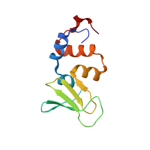
Structure of the zinc-binding domain of Bacillus stearothermophilus DNA primase.

Pan, H.Wigley, D.B.

(2000) Structure 8: 231-239

  • DOI: https://doi.org/10.1016/s0969-2126(00)00101-5
  • Primary Citation of Related Structures:  
    1D0Q

  • PubMed Abstract: 

    DNA primases catalyse the synthesis of the short RNA primers that are required for DNA replication by DNA polymerases. Primases comprise three functional domains: a zinc-binding domain that is responsible for template recognition, a polymerase domain, and a domain that interacts with the replicative helicase, DnaB. We present the crystal structure of the zinc-binding domain of DNA primase from Bacillus stearothermophilus, determined at 1.7 A resolution. This is the first high-resolution structural information about any DNA primase. A model is discussed for the interaction of this domain with the single-stranded DNA template. The structure of the DNA primase zinc-binding domain confirms that the protein belongs to the zinc ribbon subfamily. Structural comparison with other nucleic acid binding proteins suggests that the beta sheet of primase is likely to be the DNA-binding surface, with conserved residues on this surface being involved in the binding and recognition of DNA.


  • Organizational Affiliation

    Sir William Dunn School of Pathology, University of Oxford, Oxford, OX1 3RE, UK.


Macromolecules
Find similar proteins by:  (by identity cutoff)  |  3D Structure
Entity ID: 1
MoleculeChains Sequence LengthOrganismDetailsImage
DNA PRIMASE
A, B
103Geobacillus stearothermophilusMutation(s): 0 
EC: 2.7.7
UniProt
Find proteins for Q9X4D0 (Geobacillus stearothermophilus)
Explore Q9X4D0 
Go to UniProtKB:  Q9X4D0
Entity Groups  
Sequence Clusters30% Identity50% Identity70% Identity90% Identity95% Identity100% Identity
UniProt GroupQ9X4D0
Sequence Annotations
Expand
  • Reference Sequence
Experimental Data & Validation

Experimental Data

  • Method: X-RAY DIFFRACTION
  • Resolution: 1.71 Å
  • R-Value Free: 0.225 
  • R-Value Work: 0.204 
  • Space Group: P 1 21 1
Unit Cell:
Length ( Å )Angle ( ˚ )
a = 36.17α = 90
b = 58.745β = 91.73
c = 45.75γ = 90
Software Package:
Software NamePurpose
MLPHAREphasing
REFMACrefinement
DENZOdata reduction
SCALEPACKdata scaling

Structure Validation

View Full Validation Report


Entry History 

Deposition Data

Revision History  (Full details and data files)

  • Version 1.0: 2000-03-29
    Type: Initial release
  • Version 1.1: 2008-04-27
    Changes: Version format compliance
  • Version 1.2: 2011-07-13
    Changes: Version format compliance
  • Version 1.3: 2024-02-07
    Changes: Data collection, Database references, Derived calculations