Three-dimensional structure of the neurotoxin ATX Ia from Anemonia sulcata in aqueous solution determined by nuclear magnetic resonance spectroscopy.
Widmer, H., Billeter, M., Wuthrich, K.(1989) Proteins 6: 357-371
- PubMed: 2576133
- DOI: https://doi.org/10.1002/prot.340060403
- Primary Citation of Related Structures:
1ATX - PubMed Abstract:
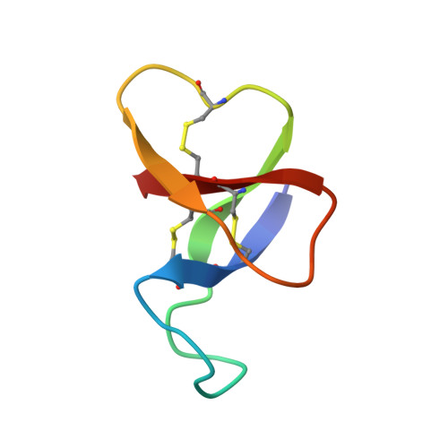
With the aid of 1H nuclear magnetic resonance (NMR) spectroscopy, the three-dimensional structure in aqueous solution was determined for ATX Ia, which is a 46 residue polypeptide neurotoxin of the sea anemone Anemonia sulcata. The input for the structure calculations consisted of 263 distance constraints from nuclear Overhauser effects (NOE) and 76 vicinal coupling constants. For the structure calculation several new or ammended programs were used in a revised strategy consisting of five successive computational steps. First, the program HABAS was used for a complete search of all backbone and chi 1 conformations that are compatible with the intraresidual and sequential NMR constraints. Second, using the program DISMAN, we extended this approach to pentapeptides by extensive sampling of all conformations that are consistent with the local and medium-range NMR constraints. Both steps resulted in the definition of additional dihedral angle constraints and in stereospecific assignments for a number of beta-methylene groups. In the next two steps DISMAN was used to obtain a group of eight conformers that contain no significant residual violations of the NMR constraints or van der Waals contacts. Finally, these structures were subjected to restrained energy refinement with a modified version of the molecular mechanics module of AMBER, which in addition to the energy force field includes potentials for the NOE distance constraints and the dihedral angle constraints. The average of the pairwise minimal RMS distances between the resulting refined conformers calculated for the well defined molecular core, which contains the backbone atoms of 35 residues and 20 interior side chains, is 1.5 +/- 0.3 A. This core is formed by a four-stranded beta-sheet connected by two well-defined loops, and there is an additional flexible loop consisting of the eleven residues 8-18. The core of the protein is stabilized by three disulfide bridges, which are surrounded by hydrophobic residues and shielded on one side by hydrophilic residues.
Organizational Affiliation:
Institut für Molekularbiologie und Biophysik, Eidgenössische Technische Hochschule-Hönggerberg, Zürich, Switzerland.