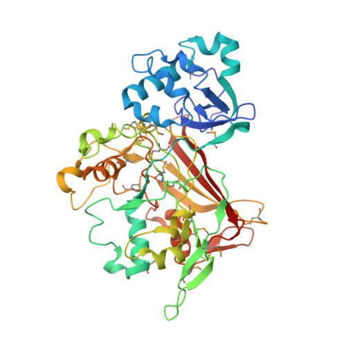
Crystal structure and mechanism of a carbon-carbon bond hydrolase.
Timm, D.E., Mueller, H.A., Bhanumoorthy, P., Harp, J.M., Bunick, G.J.(1999) Structure 7: 1023-1033
- PubMed: 10508789
- DOI: https://doi.org/10.1016/s0969-2126(99)80170-1
- Primary Citation of Related Structures:
1QCN, 1QCO, 1QQJ - PubMed Abstract:
Fumarylacetoacetate hydrolase (FAH) catalyzes the final step of tyrosine and phenylalanine catabolism, the hydrolytic cleavage of a carbon-carbon bond in fumarylacetoacetate, to yield fumarate and acetoacetate. FAH has no known sequence homologs and functions by an unknown mechanism. Carbon-carbon hydrolysis reactions are essential for the human metabolism of aromatic amino acids. FAH deficiency causes the fatal metabolic disease hereditary tyrosinemia type I. Carbon-carbon bond hydrolysis is also important in the microbial metabolism of aromatic compounds as part of the global carbon cycle. The FAH crystal structure has been determined by rapid, automated analysis of multiwavelength anomalous diffraction data. The FAH polypeptide folds into a 120-residue N-terminal domain and a 300-residue C-terminal domain. The C-terminal domain defines an unusual beta-strand topology and a novel 'mixed beta-sandwich roll' structure. The structure of FAH complexed with its physiological products was also determined. This structure reveals fumarate binding near the entrance to the active site and acetoacetate binding to an octahedrally coordinated calcium ion located in close proximity to a Glu-His dyad. FAH represents the first structure of a hydrolase that acts specifically on carbon-carbon bonds. FAH also defines a new class of metalloenzymes characterized by a unique alpha/beta fold. A mechanism involving a Glu-His-water catalytic triad is suggested based on structural observations, sequence conservation and mutational analysis. The histidine imidazole group is proposed to function as a general base. The Ca(2+) is proposed to function in binding substrate, activating the nucleophile and stabilizing a carbanion leaving group. An oxyanion hole formed from sidechains is proposed to stabilize a tetrahedral alkoxide transition state. The proton transferred to the carbanion leaving group is proposed to originate from a lysine sidechain. The results also reveal the molecular basis for mutations causing the hereditary tyrosinemia type 1.
Organizational Affiliation:
Department of Biochemistry and Molecular Biology Indiana University School of Medicine 635 Barnhill Drive, Indianapolis, Indiana 46202, USA. dtimm@iupui.edu