1IZ3

Dimeric structure of FIH (Factor inhibiting HIF)


Experimental Data Snapshot

  • Method: X-RAY DIFFRACTION
  • Resolution: 2.80 Å
  • R-Value Free: 0.277 
  • R-Value Work: 0.230 
  • R-Value Observed: 0.236 

wwPDB Validation   3D Report Full Report


This is version 1.3 of the entry. See complete history


Literature

Structure of human FIH-1 reveals a unique active site pocket and interaction sites for HIF-1 and von Hippel-Lindau.

Lee, C.Kim, S.J.Jeong, D.G.Lee, S.M.Ryu, S.E.

(2003) J Biol Chem 278: 7558-7563

  • DOI: https://doi.org/10.1074/jbc.M210385200
  • Primary Citation of Related Structures:  
    1IZ3

  • PubMed Abstract: 

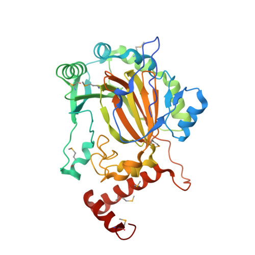
    The master switch of cellular hypoxia responses, hypoxia-inducible factor 1 (HIF-1), is hydroxylated by factor inhibiting HIF-1 (FIH-1) at a conserved asparagine residue under normoxia, which suppresses transcriptional activity of HIF-1 by abrogating its interaction with transcription coactivators. Here we report the crystal structure of human FIH-1 at 2.8-A resolution. The structural core of FIH-1 consists of a jellyroll-like beta-barrel containing the conserved ferrous-binding triad residues, confirming that FIH-1 is a member of the 2-oxoglutarate-dependent dioxygenase family. Except for the core structure and triad residues, FIH-1 has many structural deviations from other family members including N- and C-terminal insertions and various deletions in the middle of the structure. The ferrous-binding triad region is highly exposed to the solvent, which is connected to a prominent groove that may bind to a helix near the hydroxylation site of HIF-1. The structure, which is in a dimeric state, also reveals the putative von Hippel-Lindau-binding site that is distinctive to the putative HIF-1-binding site, supporting the formation of the ternary complex by FIH-1, HIF-1, and von Hippel-Lindau. The unique environment of the active site and cofactor-binding region revealed in the structure should allow design of selective drugs that can be used in ischemic diseases to promote hypoxia responses.


  • Organizational Affiliation

    Center for Cellular Switch Protein Structure, Korea Research Institute of Bioscience and Biotechnology, 52 Euh-eun-dong, Yuseong-gu, Daejeon 305-806, Korea.


Macromolecules
Find similar proteins by:  (by identity cutoff)  |  3D Structure
Entity ID: 1
MoleculeChains Sequence LengthOrganismDetailsImage
FIH349Homo sapiensMutation(s): 8 
UniProt & NIH Common Fund Data Resources
Find proteins for Q9NWT6 (Homo sapiens)
Explore Q9NWT6 
Go to UniProtKB:  Q9NWT6
PHAROS:  Q9NWT6
GTEx:  ENSG00000166135 
Entity Groups  
Sequence Clusters30% Identity50% Identity70% Identity90% Identity95% Identity100% Identity
UniProt GroupQ9NWT6
Sequence Annotations
Expand
  • Reference Sequence
Small Molecules
Ligands 1 Unique
IDChains Name / Formula / InChI Key2D Diagram3D Interactions
SO4
Query on SO4

Download Ideal Coordinates CCD File 
B [auth A]SULFATE ION
O4 S
QAOWNCQODCNURD-UHFFFAOYSA-L
Modified Residues  1 Unique
IDChains TypeFormula2D DiagramParent
MSE
Query on MSE
A
L-PEPTIDE LINKINGC5 H11 N O2 SeMET
Experimental Data & Validation

Experimental Data

  • Method: X-RAY DIFFRACTION
  • Resolution: 2.80 Å
  • R-Value Free: 0.277 
  • R-Value Work: 0.230 
  • R-Value Observed: 0.236 
  • Space Group: P 41 21 2
Unit Cell:
Length ( Å )Angle ( ˚ )
a = 86.89α = 90
b = 86.89β = 90
c = 143.42γ = 90
Software Package:
Software NamePurpose
DENZOdata reduction
SCALEPACKdata scaling
SHARPphasing
CNSrefinement

Structure Validation

View Full Validation Report


Entry History 

Deposition Data

Revision History  (Full details and data files)

  • Version 1.0: 2003-06-10
    Type: Initial release
  • Version 1.1: 2008-04-27
    Changes: Version format compliance
  • Version 1.2: 2011-07-13
    Changes: Derived calculations, Version format compliance
  • Version 1.3: 2023-12-27
    Changes: Data collection, Database references, Derived calculations