6ZOZ

Structure of Disulphide-stabilized SARS-CoV-2 Spike Protein Trimer (x1 disulphide-bond mutant, S383C, D985C, K986P, V987P, single Arg S1/S2 cleavage site) in Locked State


Experimental Data Snapshot

  • Method: ELECTRON MICROSCOPY
  • Resolution: 3.50 Å
  • Aggregation State: PARTICLE 
  • Reconstruction Method: SINGLE PARTICLE 

wwPDB Validation   3D Report Full Report


This is version 5.0 of the entry. See complete history


Literature

A thermostable, closed SARS-CoV-2 spike protein trimer.

Xiong, X.Qu, K.Ciazynska, K.A.Hosmillo, M.Carter, A.P.Ebrahimi, S.Ke, Z.Scheres, S.H.W.Bergamaschi, L.Grice, G.L.Zhang, Y.Nathan, J.A.Baker, S.James, L.C.Baxendale, H.E.Goodfellow, I.Doffinger, R.Briggs, J.A.G.

(2020) Nat Struct Mol Biol 27: 934-941

  • DOI: https://doi.org/10.1038/s41594-020-0478-5
  • Primary Citation of Related Structures:  
    6ZOX, 6ZOY, 6ZOZ, 6ZP0, 6ZP1, 6ZP2

  • PubMed Abstract: 

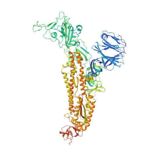
    The spike (S) protein of SARS-CoV-2 mediates receptor binding and cell entry and is the dominant target of the immune system. It exhibits substantial conformational flexibility. It transitions from closed to open conformations to expose its receptor-binding site and, subsequently, from prefusion to postfusion conformations to mediate fusion of viral and cellular membranes. S-protein derivatives are components of vaccine candidates and diagnostic assays, as well as tools for research into the biology and immunology of SARS-CoV-2. Here we have designed mutations in S that allow the production of thermostable, disulfide-bonded S-protein trimers that are trapped in the closed, prefusion state. Structures of the disulfide-stabilized and non-disulfide-stabilized proteins reveal distinct closed and locked conformations of the S trimer. We demonstrate that the designed, thermostable, closed S trimer can be used in serological assays. This protein has potential applications as a reagent for serology, virology and as an immunogen.


  • Organizational Affiliation

    Structural Studies Division, Medical Research Council Laboratory of Molecular Biology, Cambridge, UK. xiong@mrc-lmb.cam.ac.uk.


Macromolecules
Find similar proteins by:  (by identity cutoff)  |  3D Structure
Entity ID: 1
MoleculeChains Sequence LengthOrganismDetailsImage
Spike glycoprotein
A, B, C
1,247Severe acute respiratory syndrome coronavirus 2Mutation(s): 4 
Gene Names: S2
UniProt
Find proteins for P0DTC2 (Severe acute respiratory syndrome coronavirus 2)
Explore P0DTC2 
Go to UniProtKB:  P0DTC2
Entity Groups  
Sequence Clusters30% Identity50% Identity70% Identity90% Identity95% Identity100% Identity
UniProt GroupP0DTC2
Sequence Annotations
Expand
  • Reference Sequence
Oligosaccharides

Help

Entity ID: 2
MoleculeChains Length2D Diagram Glycosylation3D Interactions
2-acetamido-2-deoxy-beta-D-glucopyranose-(1-4)-2-acetamido-2-deoxy-beta-D-glucopyranose
D, E, F, G, H
D, E, F, G, H, I, J, K, L, M, N, O
2N-Glycosylation
Glycosylation Resources
GlyTouCan:  G42666HT
GlyCosmos:  G42666HT
GlyGen:  G42666HT
Small Molecules
Ligands 2 Unique
IDChains Name / Formula / InChI Key2D Diagram3D Interactions
BLA
Query on BLA

Download Ideal Coordinates CCD File 
CA [auth A],
EB [auth C],
QA [auth B]
BILIVERDINE IX ALPHA
C33 H34 N4 O6
GWZYPXHJIZCRAJ-SRVCBVSDSA-N
NAG
Query on NAG

Download Ideal Coordinates CCD File 
AA [auth A]
AB [auth C]
BA [auth A]
BB [auth C]
CB [auth C]
AA [auth A],
AB [auth C],
BA [auth A],
BB [auth C],
CB [auth C],
DA [auth B],
DB [auth C],
EA [auth B],
FA [auth B],
GA [auth B],
HA [auth B],
IA [auth B],
JA [auth B],
KA [auth B],
LA [auth B],
MA [auth B],
NA [auth B],
OA [auth B],
P [auth A],
PA [auth B],
Q [auth A],
R [auth A],
RA [auth C],
S [auth A],
SA [auth C],
T [auth A],
TA [auth C],
U [auth A],
UA [auth C],
V [auth A],
VA [auth C],
W [auth A],
WA [auth C],
X [auth A],
XA [auth C],
Y [auth A],
YA [auth C],
Z [auth A],
ZA [auth C]
2-acetamido-2-deoxy-beta-D-glucopyranose
C8 H15 N O6
OVRNDRQMDRJTHS-FMDGEEDCSA-N
Experimental Data & Validation

Experimental Data

  • Method: ELECTRON MICROSCOPY
  • Resolution: 3.50 Å
  • Aggregation State: PARTICLE 
  • Reconstruction Method: SINGLE PARTICLE 
EM Software:
TaskSoftware PackageVersion
MODEL REFINEMENTPHENIX1.18.2
RECONSTRUCTIONRELION3.1

Structure Validation

View Full Validation Report


Entry History & Funding Information

Deposition Data


Funding OrganizationLocationGrant Number
Medical Research Council (MRC, United Kingdom)United KingdomMC_UP_1201/16
European Research Council (ERC)United KingdomERC-CoG-648432
German Research Foundation (DFG)United Kingdom240245660 - SFB1129
Medical Research Council (MRC, United Kingdom)United KingdomMC_UP_A025_1013

Revision History  (Full details and data files)

  • Version 1.0: 2020-07-22
    Type: Initial release
  • Version 2.0: 2020-07-29
    Type: Remediation
    Reason: Carbohydrate remediation
    Changes: Atomic model, Data collection, Derived calculations, Structure summary
  • Version 2.1: 2020-08-12
    Changes: Database references, Structure summary
  • Version 2.2: 2020-10-21
    Changes: Database references
  • Version 2.3: 2021-02-10
    Changes: Database references
  • Version 3.0: 2021-05-26
    Changes: Advisory, Atomic model, Database references, Derived calculations, Polymer sequence, Source and taxonomy, Structure summary
  • Version 4.0: 2021-06-02
    Changes: Advisory, Atomic model, Data collection, Database references, Derived calculations, Structure summary
  • Version 5.0: 2022-03-02
    Type: Coordinate replacement
    Reason: Model completeness
    Changes: Advisory, Atomic model, Author supporting evidence, Data collection, Database references, Derived calculations, Non-polymer description, Refinement description, Source and taxonomy, Structure summary