5CDR

2.65 structure of S.aureus DNA gyrase and artificially nicked DNA


Experimental Data Snapshot

  • Method: X-RAY DIFFRACTION
  • Resolution: 2.65 Å
  • R-Value Free: 0.210 
  • R-Value Work: 0.188 
  • R-Value Observed: 0.189 

wwPDB Validation   3D Report Full Report


This is version 1.1 of the entry. See complete history


Literature

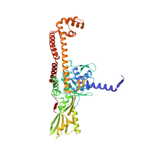
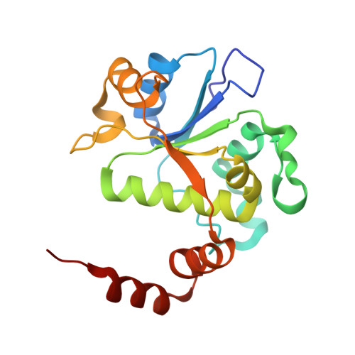
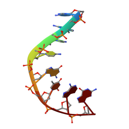
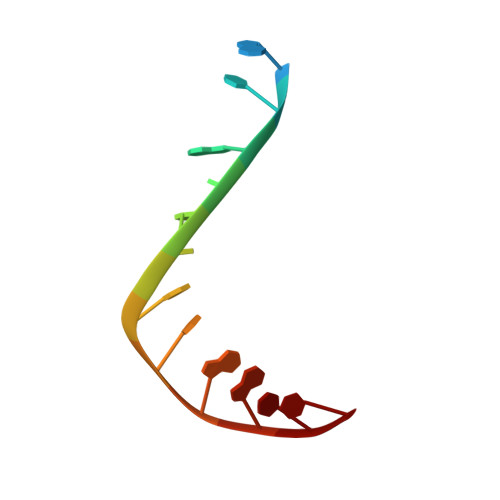
Structural basis of DNA gyrase inhibition by antibacterial QPT-1, anticancer drug etoposide and moxifloxacin.

Chan, P.F.Srikannathasan, V.Huang, J.Cui, H.Fosberry, A.P.Gu, M.Hann, M.M.Hibbs, M.Homes, P.Ingraham, K.Pizzollo, J.Shen, C.Shillings, A.J.Spitzfaden, C.E.Tanner, R.Theobald, A.J.Stavenger, R.A.Bax, B.D.Gwynn, M.N.

(2015) Nat Commun 6: 10048-10048

  • DOI: https://doi.org/10.1038/ncomms10048
  • Primary Citation of Related Structures:  
    5CDM, 5CDN, 5CDO, 5CDP, 5CDQ, 5CDR

  • PubMed Abstract: 

    New antibacterials are needed to tackle antibiotic-resistant bacteria. Type IIA topoisomerases (topo2As), the targets of fluoroquinolones, regulate DNA topology by creating transient double-strand DNA breaks. Here we report the first co-crystal structures of the antibacterial QPT-1 and the anticancer drug etoposide with Staphylococcus aureus DNA gyrase, showing binding at the same sites in the cleaved DNA as the fluoroquinolone moxifloxacin. Unlike moxifloxacin, QPT-1 and etoposide interact with conserved GyrB TOPRIM residues rationalizing why QPT-1 can overcome fluoroquinolone resistance. Our data show etoposide's antibacterial activity is due to DNA gyrase inhibition and suggests other anticancer agents act similarly. Analysis of multiple DNA gyrase co-crystal structures, including asymmetric cleavage complexes, led to a 'pair of swing-doors' hypothesis in which the movement of one DNA segment regulates cleavage and religation of the second DNA duplex. This mechanism can explain QPT-1's bacterial specificity. Structure-based strategies for developing topo2A antibacterials are suggested.


  • Organizational Affiliation

    Antibacterial Discovery Performance Unit, Infectious Diseases, Therapy Area Unit, GlaxoSmithKline, 1250 South Collegeville Road, Collegeville, Pennsylvania 19426-0989, USA.


Macromolecules

Find similar proteins by:  (by identity cutoff)  |  3D Structure
Entity ID: 1
MoleculeChains Sequence LengthOrganismDetailsImage
DNA gyrase subunit A
A, C
483Staphylococcus aureus subsp. aureus N315Mutation(s): 1 
Gene Names: gyrASA0006
EC: 5.99.1.3
UniProt
Find proteins for Q99XG5 (Staphylococcus aureus (strain N315))
Explore Q99XG5 
Go to UniProtKB:  Q99XG5
Entity Groups  
Sequence Clusters30% Identity50% Identity70% Identity90% Identity95% Identity100% Identity
UniProt GroupQ99XG5
Sequence Annotations
Expand
  • Reference Sequence
Find similar proteins by:  (by identity cutoff)  |  3D Structure
Entity ID: 2
MoleculeChains Sequence LengthOrganismDetailsImage
DNA gyrase subunit B,DNA gyrase subunit B
B, D
190Staphylococcus aureus subsp. aureus N315Mutation(s): 0 
Gene Names: gyrBSA0005
EC: 5.99.1.3
UniProt
Find proteins for P66937 (Staphylococcus aureus (strain N315))
Explore P66937 
Go to UniProtKB:  P66937
Entity Groups  
Sequence Clusters30% Identity50% Identity70% Identity90% Identity95% Identity100% Identity
UniProt GroupP66937
Sequence Annotations
Expand
  • Reference Sequence

Find similar nucleic acids by:  Sequence   |   3D Structure  

Entity ID: 3
MoleculeChains LengthOrganismImage
DNA (5'-D(*AP*GP*CP*CP*GP*TP*AP*)-3')8synthetic construct
Sequence Annotations
Expand
  • Reference Sequence

Find similar nucleic acids by:  Sequence   |   3D Structure  

Entity ID: 4
MoleculeChains LengthOrganismImage
DNA (5'-D(*GP*GP*TP*AP*CP*CP*TP*AP*CP*GP*GP*CP*T)-3')F [auth G]13synthetic construct
Sequence Annotations
Expand
  • Reference Sequence

Find similar nucleic acids by:  Sequence   |   3D Structure  

Entity ID: 5
MoleculeChains LengthOrganismImage
DNA (5'-D(*AP*GP*CP*CP*GP*TP*AP*GP*GP*TP*AP*CP*CP*TP*AP*CP*GP*GP*CP*T)-3')G [auth F]20synthetic construct
Sequence Annotations
Expand
  • Reference Sequence
Experimental Data & Validation

Experimental Data

  • Method: X-RAY DIFFRACTION
  • Resolution: 2.65 Å
  • R-Value Free: 0.210 
  • R-Value Work: 0.188 
  • R-Value Observed: 0.189 
  • Space Group: P 61
Unit Cell:
Length ( Å )Angle ( ˚ )
a = 93.395α = 90
b = 93.395β = 90
c = 410.583γ = 120
Software Package:
Software NamePurpose
SCALEPACKdata scaling
BUSTER-TNTrefinement
PDB_EXTRACTdata extraction
REFMACphasing

Structure Validation

View Full Validation Report


Entry History 

Deposition Data

Revision History  (Full details and data files)

  • Version 1.0: 2015-12-16
    Type: Initial release
  • Version 1.1: 2024-01-10
    Changes: Data collection, Database references, Derived calculations, Refinement description