Nuclear magnetic resonance structure of the nucleic acid-binding domain of severe acute respiratory syndrome coronavirus nonstructural protein 3.
Serrano, P., Johnson, M.A., Chatterjee, A., Neuman, B.W., Joseph, J.S., Buchmeier, M.J., Kuhn, P., Wuthrich, K.(2009) J Virol 83: 12998-13008
- PubMed: 19828617
- DOI: https://doi.org/10.1128/JVI.01253-09
- Primary Citation of Related Structures:
2K87 - PubMed Abstract:
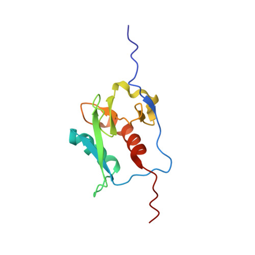
The nuclear magnetic resonance (NMR) structure of a globular domain of residues 1071 to 1178 within the previously annotated nucleic acid-binding region (NAB) of severe acute respiratory syndrome coronavirus nonstructural protein 3 (nsp3) has been determined, and N- and C-terminally adjoining polypeptide segments of 37 and 25 residues, respectively, have been shown to form flexibly extended linkers to the preceding globular domain and to the following, as yet uncharacterized domain. This extension of the structural coverage of nsp3 was obtained from NMR studies with an nsp3 construct comprising residues 1066 to 1181 [nsp3(1066-1181)] and the constructs nsp3(1066-1203) and nsp3(1035-1181). A search of the protein structure database indicates that the globular domain of the NAB represents a new fold, with a parallel four-strand beta-sheet holding two alpha-helices of three and four turns that are oriented antiparallel to the beta-strands. Two antiparallel two-strand beta-sheets and two 3(10)-helices are anchored against the surface of this barrel-like molecular core. Chemical shift changes upon the addition of single-stranded RNAs (ssRNAs) identified a group of residues that form a positively charged patch on the protein surface as the binding site responsible for the previously reported affinity for nucleic acids. This binding site is similar to the ssRNA-binding site of the sterile alpha motif domain of the Saccharomyces cerevisiae Vts1p protein, although the two proteins do not share a common globular fold.
Organizational Affiliation:
Department of Molecular Biology, The Scripps Research Institute, 10550 North Torrey Pines Rd., MB-44, La Jolla, CA 92037, USA.