2D45

Crystal structure of the MecI-mecA repressor-operator complex


Experimental Data Snapshot

  • Method: X-RAY DIFFRACTION
  • Resolution: 3.80 Å
  • R-Value Free: 0.297 
  • R-Value Work: 0.264 
  • R-Value Observed: 0.267 

wwPDB Validation   3D Report Full Report


This is version 1.4 of the entry. See complete history


Literature

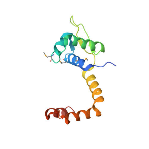
Structure of the MecI repressor from Staphylococcus aureus in complex with the cognate DNA operator of mec.

Safo, M.K.Ko, T.P.Musayev, F.N.Zhao, Q.Wang, A.H.Archer, G.L.

(2006) Acta Crystallogr Sect F Struct Biol Cryst Commun 62: 320-324

  • DOI: https://doi.org/10.1107/S1744309106009742
  • Primary Citation of Related Structures:  
    2D45

  • PubMed Abstract: 

    The dimeric repressor MecI regulates the mecA gene that encodes the penicillin-binding protein PBP-2a in methicillin-resistant Staphylococcus aureus (MRSA). MecI is similar to BlaI, the repressor for the blaZ gene of beta-lactamase. MecI and BlaI can bind to both operator DNA sequences. The crystal structure of MecI in complex with the 32 base-pair cognate DNA of mec was determined to 3.8 A resolution. MecI is a homodimer and each monomer consists of a compact N-terminal winged-helix domain, which binds to DNA, and a loosely packed C-terminal helical domain, which intertwines with its counter-monomer. The crystal contains horizontal layers of virtual DNA double helices extending in three directions, which are separated by perpendicular DNA segments. Each DNA segment is bound to two MecI dimers. Similar to the BlaI-mec complex, but unlike the MecI-bla complex, the MecI repressors bind to both sides of the mec DNA dyad that contains four conserved sequences of TACA/TGTA. The results confirm the up-and-down binding to the mec operator, which may account for cooperative effect of the repressor.


  • Organizational Affiliation

    Department of Medicinal Chemistry, School of Pharmacy and Institute for Structural Biology and Drug Discovery, Virginia Commonwealth University, Richmond, Virginia 23298, USA. msafo@vcu.edu


Macromolecules

Find similar proteins by:  (by identity cutoff)  |  3D Structure
Entity ID: 2
MoleculeChains Sequence LengthOrganismDetailsImage
Methicillin resistance regulatory protein mecIE [auth A],
F [auth B],
G [auth C],
H [auth D]
123Staphylococcus aureusMutation(s): 4 
Gene Names: mecI
UniProt
Find proteins for P68261 (Staphylococcus aureus (strain N315))
Explore P68261 
Go to UniProtKB:  P68261
Entity Groups  
Sequence Clusters30% Identity50% Identity70% Identity90% Identity95% Identity100% Identity
UniProt GroupP68261
Sequence Annotations
Expand
  • Reference Sequence

Find similar nucleic acids by:  Sequence   |   3D Structure  

Entity ID: 1
MoleculeChains LengthOrganismImage
5'-D(P*TP*AP*CP*TP*AP*CP*AP*TP*AP*TP*GP*TP*AP*GP*TP*A)-3'A [auth E],
B [auth F],
C [auth G],
D [auth H]
16N/A
Sequence Annotations
Expand
  • Reference Sequence
Small Molecules
Modified Residues  1 Unique
IDChains TypeFormula2D DiagramParent
MSE
Query on MSE
E [auth A],
F [auth B],
G [auth C],
H [auth D]
L-PEPTIDE LINKINGC5 H11 N O2 SeMET
Experimental Data & Validation

Experimental Data

  • Method: X-RAY DIFFRACTION
  • Resolution: 3.80 Å
  • R-Value Free: 0.297 
  • R-Value Work: 0.264 
  • R-Value Observed: 0.267 
  • Space Group: P 32 1 2
Unit Cell:
Length ( Å )Angle ( ˚ )
a = 61.464α = 90
b = 61.464β = 90
c = 419.777γ = 120
Software Package:
Software NamePurpose
HKL-2000data collection
SCALEPACKdata scaling
AMoREphasing
CNSrefinement
HKL-2000data reduction

Structure Validation

View Full Validation Report


Entry History 

Deposition Data

Revision History  (Full details and data files)

  • Version 1.0: 2005-10-25
    Type: Initial release
  • Version 1.1: 2008-04-30
    Changes: Version format compliance
  • Version 1.2: 2011-07-13
    Changes: Version format compliance
  • Version 1.3: 2023-10-25
    Changes: Data collection, Database references, Derived calculations, Refinement description
  • Version 1.4: 2023-11-15
    Changes: Data collection