Structural and functional insights into the interaction between a PP01 phage gp38 tail fiber tip and an Escherichia coli OmpC receptor.
Terasaki, H., Zdravkovic, A., Niwa, T., Washizaki, A., Kawaguchi, M., Yonesaki, T., Kanamaru, S., Otsuka, Y.(2026) mBio : e0211025-e0211025
- PubMed: 41511108
- DOI: https://doi.org/10.1128/mbio.02110-25
- Primary Citation of Related Structures:
9V85 - PubMed Abstract:
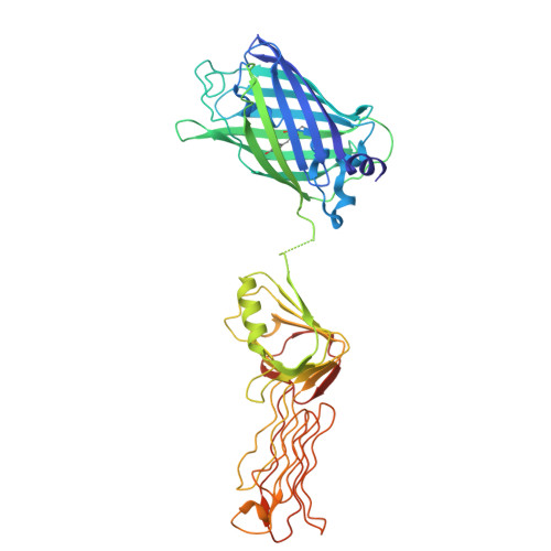
Bacteriophages exhibit strict host specificity, primarily determined by adsorption to bacterial surface receptors. However, the molecular basis underlying this specificity remains incompletely understood. Here, we investigate the interaction between outer membrane protein C (OmpC) of Escherichia coli O157 and gp38, the receptor-binding protein located at the tip of the long tail fibers of phage PP01. We determined the crystal structure of the receptor-binding domain (RBD) of gp38 PP01 at 2.1 Å resolution. The structure reveals a lattice of poly-glycine type II helices with protruding receptor recognition loops, resembling that of gp38 from Salmonella phage S16. To identify interaction sites, we performed site-specific photo-crosslinking using p -benzoyl-L-phenylalanine (pBPA), followed by liquid chromatography-tandem mass spectrometry. Two critical contacts were identified: Gly208 in loop-D of gp38 PP01 crosslinked to Ser225 and Pro226 in extracellular loop-5 of OmpC O157 , and Tyr230 in loop-E of gp38 PP01 to the Val304-Arg308 region in loop-7 of OmpC O157 . A structural model of the gp38 PP01 -OmpC O157 complex was constructed using distance-constrained prediction and validated by targeted mutagenesis. Our findings demonstrate that PP01 phage specificity is governed by loop-E of gp38 PP01 engaging a cleft formed by loops -5 and -7 of OmpC O157 . These structural and functional insights enhance our understanding of phage-host recognition and may inform the rational design of engineered bacteriophages with altered host ranges.IMPORTANCEBacteriophages must precisely recognize and bind to specific molecules on the surface of their bacterial hosts to initiate infection, but the details of these interactions are often unclear. In this study, we examined how phage PP01 targets Escherichia coli O157. Using structural analysis of the phage tail fiber and a technique to capture contact points between the phage and a bacterial surface protein, we mapped the molecular basis of host recognition. We also developed a simple test system using a modified phage to identify which parts of the tail fiber are essential for binding. These methods can be broadly applied to other phages to better understand how they select their hosts. This work provides valuable insights and tools that could aid the design of phages with customized host specificity for therapeutic or biotechnological applications.
- Department of Biochemistry and Molecular Biology, Graduate School of Science and Engineering, Saitama University, Saitama, Japan.
Organizational Affiliation: