9SIN | pdb_00009sin

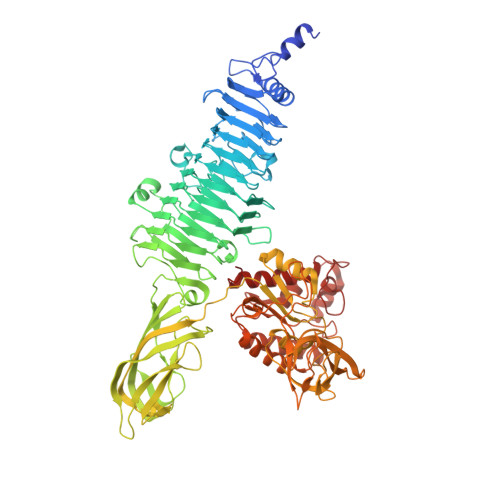
Phage epsilon15 tailspike gp20 containing domains beta-helix, beta-sandwich and petal domains with three hexasaccharides of the Salmonella Anatum O-antigen


Experimental Data Snapshot

  • Method: X-RAY DIFFRACTION
  • Resolution: 2.00 Å
  • R-Value Free: 
    0.191 (Depositor), 0.191 (DCC) 
  • R-Value Work: 
    0.166 (Depositor), 0.167 (DCC) 

Starting Model: experimental
View more details

wwPDB Validation 3D Report Full Report

Validation slider image for 9SIN

This is version 1.0 of the entry. See complete history

Literature

Structure of the Salmonella bacteriophage epsilon15 tailspike reveals multiple lipopolysaccharide binding sites and protruding esterase domains.

Seoane-Blanco, M.Pereda, A.Broeker, N.McConnell, M.Canada, F.J.Barbirz, S.van Raaij, M.J.

To be published.

Macromolecule Content 

  • Total Structure Weight: 96.01 kDa 
  • Atom Count: 7,265 
  • Modeled Residue Count: 832 
  • Deposited Residue Count: 857 
  • Unique protein chains: 1

Macromolecules

Find similar proteins by:|  3D Structure
Entity ID: 1
MoleculeChains  Sequence LengthOrganismDetailsImage
Tail spike protein857Salmonella phage epsilon15Mutation(s): 0 
EC: 3.2.1
UniProt
Find proteins for Q858F5 (Salmonella phage epsilon15)
Explore Q858F5 
Go to UniProtKB:  Q858F5
Entity Groups
Sequence Clusters30% Identity50% Identity70% Identity90% Identity95% Identity100% Identity
UniProt GroupQ858F5
Sequence Annotations
Expand
Reference Sequence

Oligosaccharides

Help  
Entity ID: 2
MoleculeChains Length2D Diagram GlycosylationD Interactions
6-O-acetyl-alpha-D-galactopyranose-(1-6)-beta-D-mannopyranose-(1-4)-alpha-L-rhamnopyranose-(1-3)-6-O-acetyl-alpha-D-galactopyranose-(1-6)-beta-D-mannopyranose-(1-4)-alpha-L-rhamnopyranose
B
6N/AN/A
Entity ID: 3
MoleculeChains Length2D Diagram GlycosylationD Interactions
alpha-D-galactopyranose-(1-6)-beta-D-mannopyranose-(1-4)-alpha-L-rhamnopyranose-(1-3)-alpha-D-galactopyranose-(1-6)-beta-D-mannopyranose-(1-4)-alpha-L-rhamnopyranoseC [auth D],
D [auth E]
6N/A

Small Molecules

Ligands 2 Unique
IDChains Name / Formula / InChI Key2D Diagram3D Interactions
FMT

Query on FMT


Download:Ideal Coordinates CCD File
E [auth A]
F [auth A]
G [auth A]
H [auth A]
I [auth A]
E [auth A],
F [auth A],
G [auth A],
H [auth A],
I [auth A],
J [auth A],
K [auth A],
L [auth A],
M [auth A],
N [auth A],
O [auth A],
P [auth A],
Q [auth A],
R [auth A],
S [auth A]
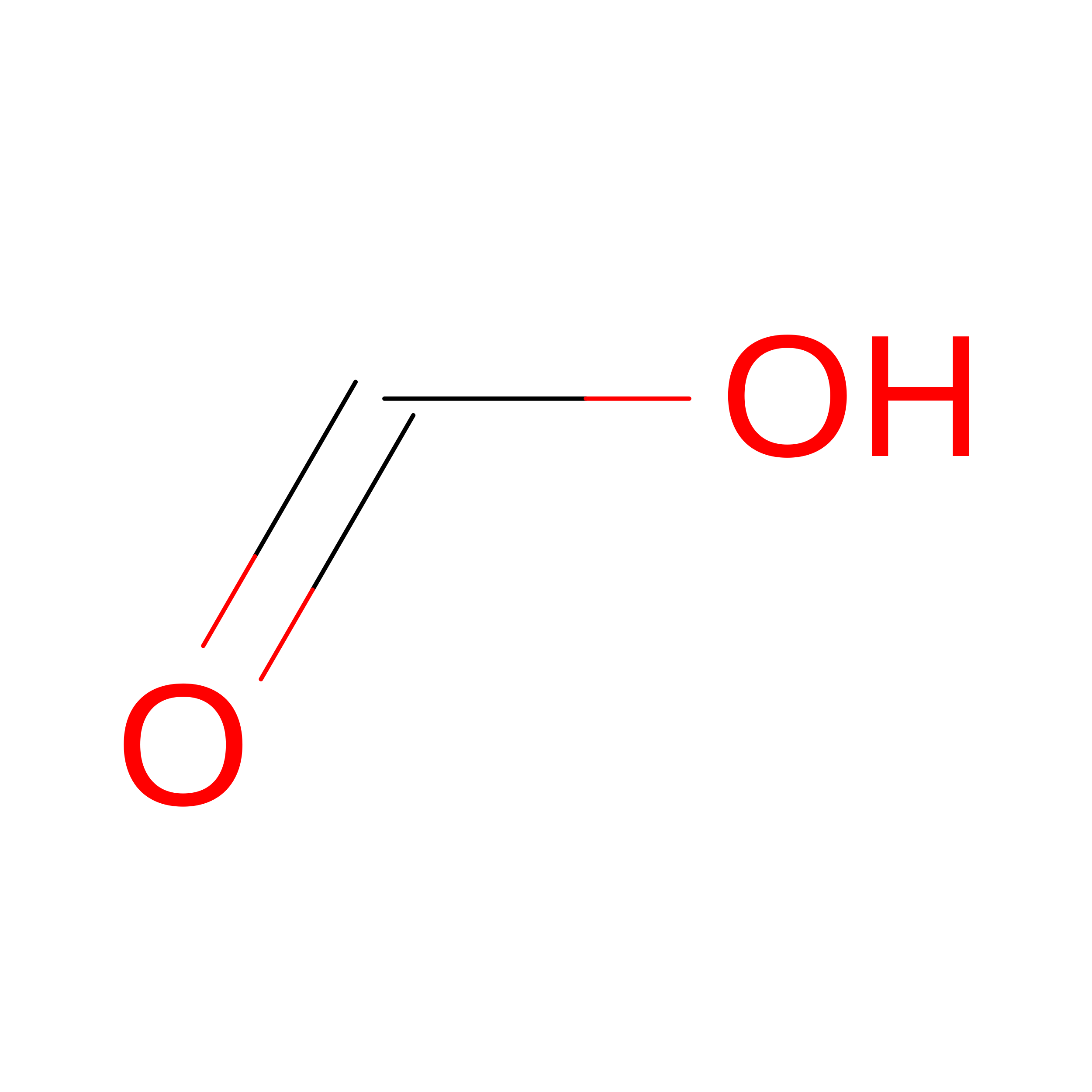
FORMIC ACID
C H2 O2
BDAGIHXWWSANSR-UHFFFAOYSA-N
NA

Query on NA


Download:Ideal Coordinates CCD File
T [auth A],
U [auth A]
SODIUM ION
Na
FKNQFGJONOIPTF-UHFFFAOYSA-N

Experimental Data & Validation

Experimental Data

  • Method: X-RAY DIFFRACTION
  • Resolution: 2.00 Å
  • R-Value Free:  0.191 (Depositor), 0.191 (DCC) 
  • R-Value Work:  0.166 (Depositor), 0.167 (DCC) 
Space Group: H 3
Unit Cell:
Length ( Å )Angle ( ˚ )
a = 117.522α = 90
b = 117.522β = 90
c = 250.68γ = 120
Software Package:
Software NamePurpose
REFMACrefinement
XDSdata reduction
Aimlessdata scaling
MOLREPphasing

Structure Validation

View Full Validation Report


Entry History 

& Funding Information

Deposition Data


Funding OrganizationLocationGrant Number
Ministerio de Ciencia e Innovacion (MCIN)SpainPID2021-125597NB-I00
Ministerio de Ciencia e Innovacion (MCIN)SpainBFU2017-82207-P
Ministerio de Ciencia e Innovacion (MCIN)SpainBFU2014-53425-P
Ministerio de Ciencia e Innovacion (MCIN)SpainBFU2011-24843/BMC

Revision History  (Full details and data files)

  • Version 1.0: 2026-09-09
    Type: Initial release