


M18BP1 valency and a distributed interaction footprint determine epigenetic centromere specification in humans
Walstein, K., Hill, L., Vetter, I.R., Pan, D., Musacchio, A.(2026) EMBO J
Experimental Data Snapshot
Starting Model: experimental
View more details
wwPDB Validation 3D Report Full Report

(2026) EMBO J
Entity ID: 1 | |||||
|---|---|---|---|---|---|
| Molecule | Chains | Sequence Length | Organism | Details | Image |
| Genome polyprotein,Mis18-binding protein 1,Chains: A | 487 | Homo sapiens | Mutation(s): 1 EC: 3.4.22.29 (PDB Primary Data), 3.6.1.15 (PDB Primary Data), 3.4.22.28 (PDB Primary Data), 2.7.7.48 (PDB Primary Data) | 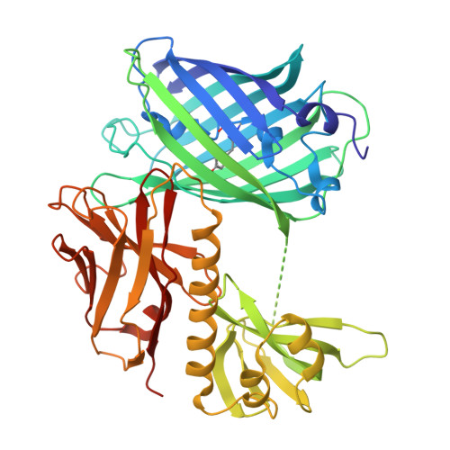 | |
UniProt & NIH Common Fund Data Resources | |||||
Find proteins for B6F2F5 (Human enterovirus 71) Explore B6F2F5 Go to UniProtKB: B6F2F5 | |||||
Find proteins for Q6P0N0 (Homo sapiens) Explore Q6P0N0 Go to UniProtKB: Q6P0N0 | |||||
PHAROS: Q6P0N0 GTEx: ENSG00000129534 | |||||
Entity Groups | |||||
| Sequence Clusters | 30% Identity50% Identity70% Identity90% Identity95% Identity100% Identity | ||||
| UniProt Groups | B6F2F5Q6P0N0 | ||||
Sequence AnnotationsExpand | |||||
| |||||
| Ligands 1 Unique | |||||
|---|---|---|---|---|---|
| ID | Chains | Name / Formula / InChI Key | 2D Diagram | 3D Interactions | |
| PG4 Query on PG4 | C [auth A] D [auth A] E [auth A] F [auth A] G [auth A] | TETRAETHYLENE GLYCOL C8 H18 O5 UWHCKJMYHZGTIT-UHFFFAOYSA-N |  | ||
| Modified Residues 1 Unique | |||||
|---|---|---|---|---|---|
| ID | Chains | Type | Formula | 2D Diagram | Parent |
| C12 Query on C12 | A, B | L-PEPTIDE LINKING | C15 H18 N3 O5 |  | THR, TYR, GLY |
| Length ( Å ) | Angle ( ˚ ) |
|---|---|
| a = 143.2 | α = 90 |
| b = 120.28 | β = 121.28 |
| c = 77.36 | γ = 90 |
| Software Name | Purpose |
|---|---|
| PHENIX | refinement |
| XSCALE | data scaling |
| PDB_EXTRACT | data extraction |
| XDS | data reduction |
| PHASER | phasing |
| Funding Organization | Location | Grant Number |
|---|---|---|
| European Research Council (ERC) | European Union | 951430 |
| German Research Foundation (DFG) | Germany | CCR 1430 |


