9PW2 | pdb_00009pw2

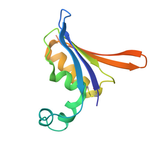
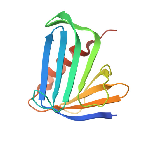
Taylorella equigenitalis BaDTF3 deaminase toxin DddB bound to the immunity protein


Experimental Data Snapshot

  • Method: X-RAY DIFFRACTION
  • Resolution: 1.61 Å
  • R-Value Free: 
    0.210 (Depositor), 0.211 (DCC) 
  • R-Value Work: 
    0.185 (Depositor), 0.185 (DCC) 
  • R-Value Observed: 
    0.186 (Depositor) 

Starting Model: in silico
View more details

wwPDB Validation 3D Report Full Report

Validation slider image for 9PW2

This is version 1.1 of the entry. See complete history

Literature

Structural basis for double-stranded DNA cytosine deamination by BaDTF3 and its application in mitochondrial genome editing.

Yin, L.Lim, C.J.Shi, K.Yoon, J.H.Ko, B.K.Ryou, S.Kim, J.S.Aihara, H.

(2026) Nat Commun 17

  • DOI: https://doi.org/10.1038/s41467-026-72730-z
  • Primary Citation Related Structures: 
    9PW0, 9PW1, 9PW2

  • PubMed Abstract: 

    Bacterial deaminase toxin family (BaDTF) proteins are weapons used in bacterial warfare, and they are useful tools in base editing, epigenetics analyses, and genomic footprinting applications. Our previous studies revealed the mechanisms of 5'-TC-specific cytosine deamination in double-stranded (ds)DNA by DddA from BaDTF1 and sequence context-independent single-stranded (ss)DNA cytosine deamination by SsdA from BaDTF2. Here, we show that a representative member of BaDTF3, DddB, deaminates cytosines specifically in dsDNA, but with a broad sequence context preference. Our crystal structure of DddB bound to dsDNA reveals a distinct mechanism of substrate engagement, in which a helix-hairpin-helix motif inserted into the minor groove of dsDNA promotes flipping of the target cytosine into the enzyme active site. Based on the structural information, we generate both monomeric and split DddB-derived cytosine base editors (BdCBE) and demonstrate that they can perform CRISPR-free mitochondrial base editing in human cells, with an expanded targeting scope compared to the DddA-derived DdCBEs. Our studies highlight the mechanistic diversity among BaDTF proteins and expand the repertoire of dsDNA deaminase enzymes for genome editing and other applications.


  • Organizational Affiliation
    • Department of Biochemistry, Molecular Biology and Biophysics, University of Minnesota, Minneapolis, MN, USA.

Macromolecule Content 

  • Total Structure Weight: 27.9 kDa 
  • Atom Count: 1,984 
  • Modeled Residue Count: 220 
  • Deposited Residue Count: 239 
  • Unique protein chains: 2

Macromolecules

Find similar proteins by:|  3D Structure
Entity ID: 1
MoleculeChains  Sequence LengthOrganismDetailsImage
BaDTF3116Taylorella equigenitalisMutation(s): 0 
Entity Groups
Sequence Clusters30% Identity50% Identity70% Identity90% Identity95% Identity100% Identity
Sequence Annotations
Expand
Reference Sequence
Find similar proteins by:|  3D Structure
Entity ID: 2
MoleculeChains  Sequence LengthOrganismDetailsImage
MEI123Taylorella equigenitalisMutation(s): 0 
Entity Groups
Sequence Clusters30% Identity50% Identity70% Identity90% Identity95% Identity100% Identity
Sequence Annotations
Expand
Reference Sequence

Experimental Data & Validation

Experimental Data

  • Method: X-RAY DIFFRACTION
  • Resolution: 1.61 Å
  • R-Value Free:  0.210 (Depositor), 0.211 (DCC) 
  • R-Value Work:  0.185 (Depositor), 0.185 (DCC) 
  • R-Value Observed: 0.186 (Depositor) 
Space Group: P 21 21 2
Unit Cell:
Length ( Å )Angle ( ˚ )
a = 58.399α = 90
b = 97.256β = 90
c = 41.968γ = 90
Software Package:
Software NamePurpose
PHENIXrefinement
XDSdata reduction
Aimlessdata scaling
PHASERphasing

Structure Validation

View Full Validation Report


Entry History 

& Funding Information

Deposition Data


Funding OrganizationLocationGrant Number
National Institutes of Health/National Institute of General Medical Sciences (NIH/NIGMS)United StatesR35GM118047

Revision History  (Full details and data files)

  • Version 1.0: 2026-08-05
    Type: Initial release
  • Version 1.1: 2026-09-02
    Changes: Database references