9LJF | pdb_00009ljf

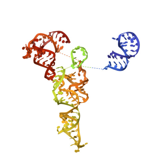
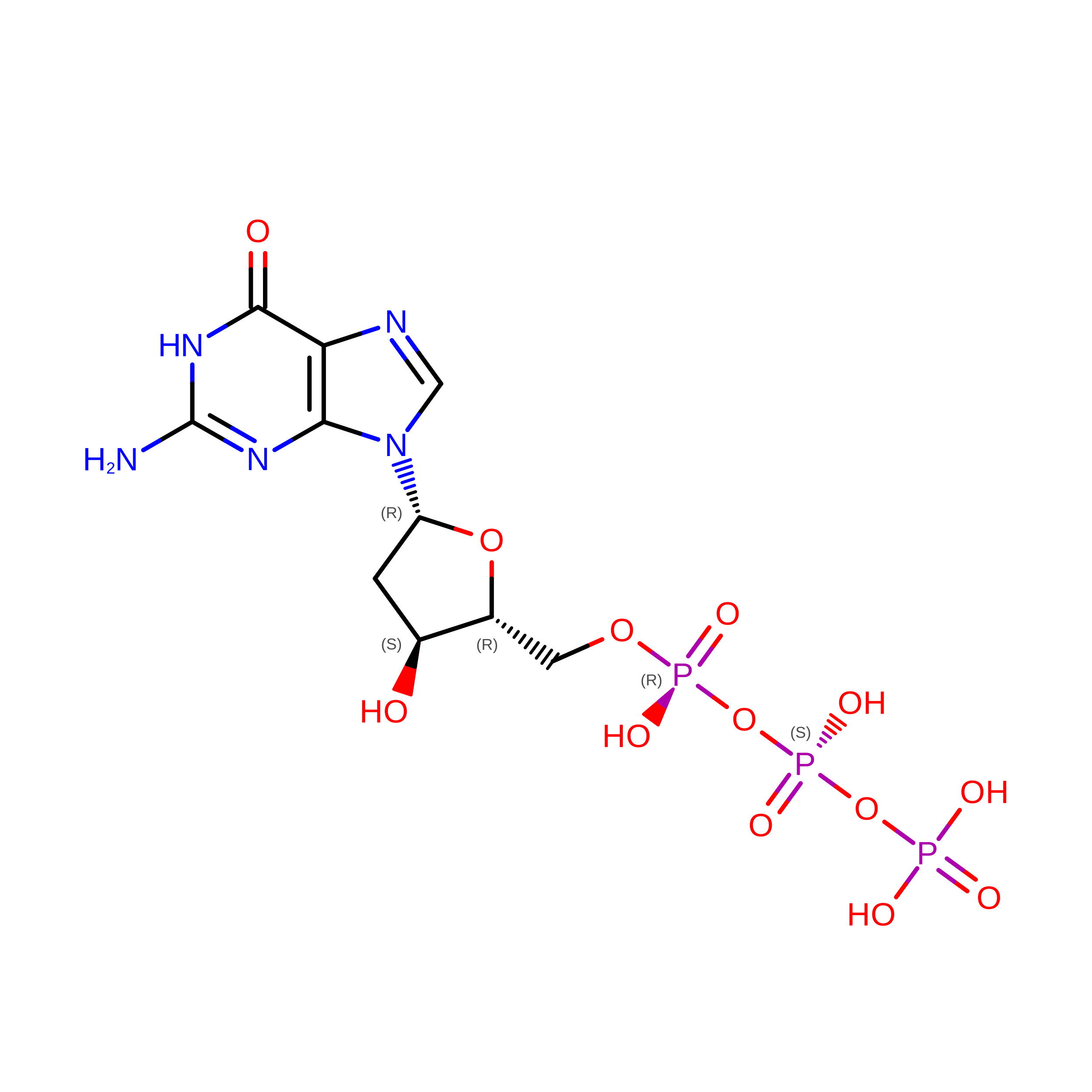
Cryo-EM structure of the dGTP bound DRT2-ncRNA complex


Experimental Data Snapshot

  • Method: ELECTRON MICROSCOPY
  • Resolution: 3.52 Å
  • Aggregation State: PARTICLE 
  • Reconstruction Method: SINGLE PARTICLE 

wwPDB Validation 3D Report Full Report

Validation slider image for 9LJF

This is version 1.1 of the entry. See complete history

Literature

Dual sensing activates antiviral reverse transcriptase for membrane targeting.

Xia, Y.Qi, H.Wei, X.Y.Zhang, J.T.Hu, W.Liu, Y.J.Song, X.Y.Liu, X.Wang, C.Jia, N.

(2026) Cell Host Microbe 

  • DOI: https://doi.org/10.1016/j.chom.2026.08.006
  • Primary Citation Related Structures: 
    9LJE, 9LJF

  • PubMed Abstract: 

    The prokaryotic type 2 defense-associated reverse transcriptase (DRT2) system mediates antiviral defense by catalyzing the rolling-circle reverse transcription of a noncoding RNA (ncRNA) and producing the toxic Neo protein that arrests bacterial growth. However, the mechanisms underlying DRT2 activation and effector function remain unknown. Here, we identified two distinct activation mechanisms: direct binding of a phage-encoded single-stranded DNA-binding protein (SSB or SSAP), and the detection of elevated intracellular dGTP levels induced by the phage-encoded ribonucleotide reductase NrdAB. Upon activation, the produced Neo protein directly targets the bacterial membrane, inducing membrane depolarization and growth arrest. Cryo-electron microscopy (cryo-EM) structures of the DRT2-ncRNA complex in its arrested and dGTP-bound active states provide mechanistic insights into rolling-circle ccDNA synthesis and template jumping. Furthermore, these identified activation mechanisms enable the DRT2 system with an engineered ncRNA template to produce a large-scale, user-defined double-stranded DNA (dsDNA) template in vivo, highlighting its potential in biotechnological applications.


  • Organizational Affiliation
    • Department of Biochemistry, SUSTech Homeostatic Medicine Institute, School of Medicine, Southern University of Science and Technology, Shenzhen 518055, China.

Macromolecule Content 

  • Total Structure Weight: 141.83 kDa 
  • Atom Count: 6,697 
  • Modeled Residue Count: 568 
  • Deposited Residue Count: 710 
  • Unique protein chains: 1
  • Unique nucleic acid chains: 2

Macromolecules


Find similar proteins by:|  3D Structure
Entity ID: 1
MoleculeChains  Sequence LengthOrganismDetailsImage
RNA-dependent DNA polymerase425Klebsiella pneumoniaeMutation(s): 0 
Entity Groups
Sequence Clusters30% Identity50% Identity70% Identity90% Identity95% Identity100% Identity
Sequence Annotations
Expand
Reference Sequence
Find similar nucleic acids by:  (by identity cutoff) 
Entity ID: 2
MoleculeChains LengthOrganismImage
RNA (280-MER)280Klebsiella pneumoniae
Sequence Annotations
Expand
Reference Sequence
Find similar nucleic acids by:  Sequence
Entity ID: 3
MoleculeChains LengthOrganismImage
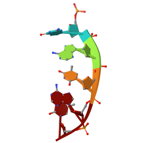
DNA (5'-D(P*GP*AP*TP*AP*T)-3')5Klebsiella pneumoniae
Sequence Annotations
Expand
Reference Sequence

Experimental Data & Validation

Experimental Data

  • Method: ELECTRON MICROSCOPY
  • Resolution: 3.52 Å
  • Aggregation State: PARTICLE 
  • Reconstruction Method: SINGLE PARTICLE 
EM Software:
TaskSoftware PackageVersion
MODEL REFINEMENTPHENIX1.20.1_4487:

Structure Validation

View Full Validation Report


Entry History 

& Funding Information

Deposition Data


Funding OrganizationLocationGrant Number
Not funded--

Revision History  (Full details and data files)

  • Version 1.0: 2026-07-15
    Type: Initial release
  • Version 1.1: 2026-09-16
    Changes: Data collection, Database references