9HPO | pdb_00009hpo

Docedameric RuvBL1/RuvBL2

  • Classification: CHAPERONE
  • Organism(s): Homo sapiens
  • Expression System: Escherichia coli
  • Mutation(s): No 

  • Deposited: 2024-12-13 Released: 2025-12-24 
  • Deposition Author(s): Santo, P.E., Plisson-Chastang, C.
  • Funding Organization(s): Centre National de la Recherche Scientifique (CNRS), Fundacao para a Ciencia e a Tecnologia, Other government

Experimental Data Snapshot

  • Method: ELECTRON MICROSCOPY
  • Resolution: 3.50 Å
  • Aggregation State: PARTICLE 
  • Reconstruction Method: SINGLE PARTICLE 

wwPDB Validation   3D Report Full Report


This is version 1.1 of the entry. See complete history


Literature

An integrative structural biology approach reveals the dynamic organization of the R2SP quaternary chaperone complex.

Santo, P.E.Chagot, M.E.Gizardin-Fredon, H.Ley, M.Chenuel, T.Desligniere, E.Plassart, L.Paiva, A.C.F.Sousa, P.M.F.Bertrand, E.Charpentier, B.Verheggen, C.Quinternet, M.Meyer, P.Bandeiras, T.M.Cianferani, S.Plisson-Chastang, C.Manival, X.

(2026) Nat Commun 

  • DOI: https://doi.org/10.1038/s41467-026-69157-x
  • Primary Citation of Related Structures:  
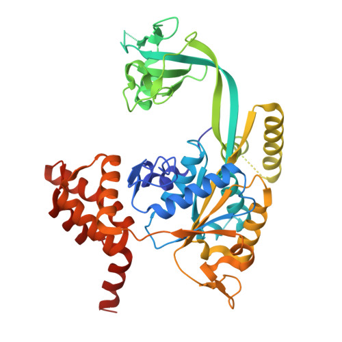
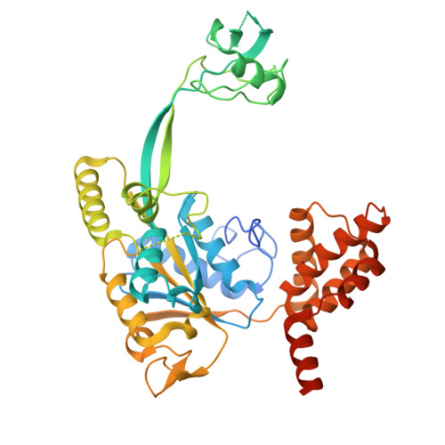
    9HB4, 9HPO

  • PubMed Abstract: 

    R2SP belongs to the R2TP-like quaternary chaperone family and consists of RUVBL1/RUVBL2 AAA+ ATPases, which powers the machinery, and SPAG1 and PIH1D2 adapter proteins that engage specific clients to promote their quaternary assembly. However, little is known about the structure of R2SP and the precise mode of action of these R2TP-like complexes. Here, we combined biochemical (ATPase and fluorescence polarization assays) and structural approaches (NMR, structural mass spectrometry, cryo-EM) to investigate the 3D organization of the R2SP complex, its mode of assembly and ATPase activity. Together with our binding, mutational and kinetic studies, these results led us to propose a model in which SPAG1 and PIH1D2 bind and cooperatively engage RUVBL1/RUVBL2 to produce R2SP. This reveals a 3D structure close to the canonical R2TP complex but also highlights differences in RUVBL1/RUVBL2 ATPase activity, as well as the cooperative binding of SPAG1 and PIH1D2 to this catalytic core that may explain functional difference between the two systems.


  • Organizational Affiliation
    • Centre de Biologie Intégrative (CBI), University of Toulouse CNRS, Equipe labellisée Ligue Nationale Contre le Cancer, Toulouse, France.

Macromolecules
Find similar proteins by:  (by identity cutoff)  |  3D Structure
Entity ID: 1
MoleculeChains Sequence LengthOrganismDetailsImage
RuvB-like 1
A, B, C, G, H
A, B, C, G, H, I
451Homo sapiensMutation(s): 0 
Gene Names: RUVBL1INO80HNMP238TIP49TIP49A
EC: 3.6.4.12
UniProt & NIH Common Fund Data Resources
Find proteins for Q9Y265 (Homo sapiens)
Explore Q9Y265 
Go to UniProtKB:  Q9Y265
PHAROS:  Q9Y265
GTEx:  ENSG00000175792 
Entity Groups  
Sequence Clusters30% Identity50% Identity70% Identity90% Identity95% Identity100% Identity
UniProt GroupQ9Y265
Sequence Annotations
Expand
  • Reference Sequence
Find similar proteins by:  (by identity cutoff)  |  3D Structure
Entity ID: 2
MoleculeChains Sequence LengthOrganismDetailsImage
RuvB-like 2
D, E, F, J, K
D, E, F, J, K, L
463Homo sapiensMutation(s): 0 
Gene Names: RUVBL2INO80JTIP48TIP49BCGI-46
EC: 3.6.4.12
UniProt & NIH Common Fund Data Resources
Find proteins for Q9Y230 (Homo sapiens)
Explore Q9Y230 
Go to UniProtKB:  Q9Y230
PHAROS:  Q9Y230
GTEx:  ENSG00000183207 
Entity Groups  
Sequence Clusters30% Identity50% Identity70% Identity90% Identity95% Identity100% Identity
UniProt GroupQ9Y230
Sequence Annotations
Expand
  • Reference Sequence
Small Molecules
Ligands 1 Unique
IDChains Name / Formula / InChI Key2D Diagram3D Interactions
ADP (Subject of Investigation/LOI)
Query on ADP

Download Ideal Coordinates CCD File 
M [auth A]
N [auth B]
O [auth C]
P [auth D]
Q [auth E]
M [auth A],
N [auth B],
O [auth C],
P [auth D],
Q [auth E],
R [auth F],
S [auth G],
T [auth H],
U [auth I],
V [auth J],
W [auth K],
X [auth L]
ADENOSINE-5'-DIPHOSPHATE
C10 H15 N5 O10 P2
XTWYTFMLZFPYCI-KQYNXXCUSA-N
Experimental Data & Validation

Experimental Data

  • Method: ELECTRON MICROSCOPY
  • Resolution: 3.50 Å
  • Aggregation State: PARTICLE 
  • Reconstruction Method: SINGLE PARTICLE 
EM Software:
TaskSoftware PackageVersion
MODEL REFINEMENTPHENIX1.21
RECONSTRUCTIONRELION3.0

Structure Validation

View Full Validation Report


Entry History & Funding Information

Deposition Data


Funding OrganizationLocationGrant Number
Centre National de la Recherche Scientifique (CNRS)FranceANR-16-CE11-0032-04
Centre National de la Recherche Scientifique (CNRS)FranceANR-23-CE12-0022 YMCR
Centre National de la Recherche Scientifique (CNRS)FranceANR-23-CE44-0035 NAProt-XLMS
Fundacao para a Ciencia e a TecnologiaPortugalUIDB/04462/2020
Fundacao para a Ciencia e a TecnologiaPortugalUIDP/04462/2020
Other governmentProFI - ANR-10-INBS-08-03
Other governmentLS4FUTURE - LA/P/0087/2020

Revision History  (Full details and data files)

  • Version 1.0: 2025-12-24
    Type: Initial release
  • Version 1.1: 2026-02-18
    Changes: Data collection, Database references