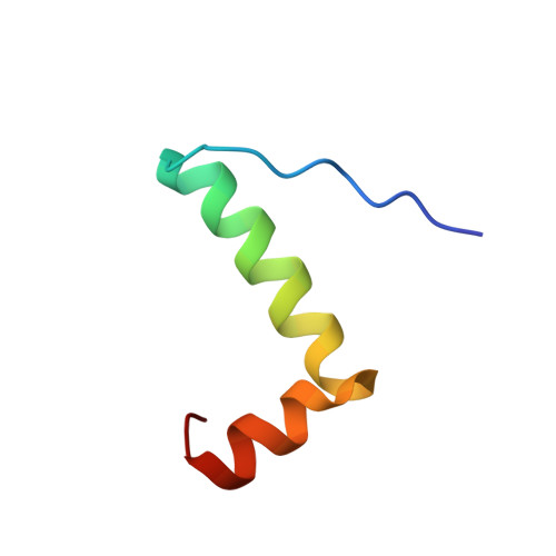
The carboxy terminus causes interfacial assembly of oleate hydratase on a membrane bilayer.
Radka, C.D., Grace, C.R., Hasdemir, H.S., Li, Y., Rodriguez, C.C., Rodrigues, P., Oldham, M.L., Qayyum, M.Z., Pitre, A., MacCain, W.J., Kalathur, R.C., Tajkhorshid, E., Rock, C.O.(2024) J Biol Chem 300: 105627-105627
- PubMed: 38211817
- DOI: https://doi.org/10.1016/j.jbc.2024.105627
- Primary Citation of Related Structures:
8UM1, 8UM2, 8UR3, 8UR6 - PubMed Abstract:
The soluble flavoprotein oleate hydratase (OhyA) hydrates the 9-cis double bond of unsaturated fatty acids. OhyA substrates are embedded in membrane bilayers; OhyA must remove the fatty acid from the bilayer and enclose it in the active site. Here, we show that the positively charged helix-turn-helix motif in the carboxy terminus (CTD) is responsible for interacting with the negatively charged phosphatidylglycerol (PG) bilayer. Super-resolution microscopy of Staphylococcus aureus cells expressing green fluorescent protein fused to OhyA or the CTD sequence shows subcellular localization along the cellular boundary, indicating OhyA is membrane-associated and the CTD sequence is sufficient for membrane recruitment. Using cryo-electron microscopy, we solved the OhyA dimer structure and conducted 3D variability analysis of the reconstructions to assess CTD flexibility. Our surface plasmon resonance experiments corroborated that OhyA binds the PG bilayer with nanomolar affinity and we found the CTD sequence has intrinsic PG binding properties. We determined that the nuclear magnetic resonance structure of a peptide containing the CTD sequence resembles the OhyA crystal structure. We observed intermolecular NOE from PG liposome protons next to the phosphate group to the CTD peptide. The addition of paramagnetic MnCl 2 indicated the CTD peptide binds the PG surface but does not insert into the bilayer. Molecular dynamics simulations, supported by site-directed mutagenesis experiments, identify key residues in the helix-turn-helix that drive membrane association. The data show that the OhyA CTD binds the phosphate layer of the PG surface to obtain bilayer-embedded unsaturated fatty acids.
Organizational Affiliation:
Department of Microbiology, Immunology, and Molecular Genetics, University of Kentucky, Lexington, Kentucky, USA; Department of Host Microbe Interactions, St Jude Children's Research Hospital, Memphis, Tennessee, USA. Electronic address: christopher.radka@uky.edu.