7WWF

Crystal structure of BioH3 from Mycolicibacterium smegmatis


Experimental Data Snapshot

  • Method: X-RAY DIFFRACTION
  • Resolution: 2.27 Å
  • R-Value Free: 0.222 
  • R-Value Work: 0.189 
  • R-Value Observed: 0.191 

wwPDB Validation   3D Report Full Report


Ligand Structure Quality Assessment 


This is version 1.1 of the entry. See complete history


Literature

Three enigmatic BioH isoenzymes are programmed in the early stage of mycobacterial biotin synthesis, an attractive anti-TB drug target.

Xu, Y.Yang, J.Li, W.Song, S.Shi, Y.Wu, L.Sun, J.Hou, M.Wang, J.Jia, X.Zhang, H.Huang, M.Lu, T.Gan, J.Feng, Y.

(2022) PLoS Pathog 18: e1010615-e1010615

  • DOI: https://doi.org/10.1371/journal.ppat.1010615
  • Primary Citation of Related Structures:  
    7WWF

  • PubMed Abstract: 

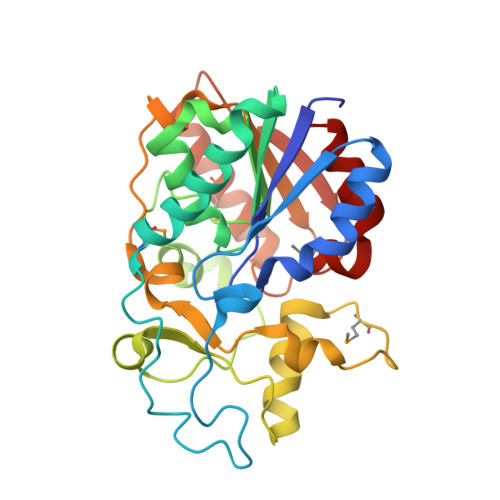
    Tuberculosis (TB) is one of the leading infectious diseases of global concern, and one quarter of the world's population are TB carriers. Biotin metabolism appears to be an attractive anti-TB drug target. However, the first-stage of mycobacterial biotin synthesis is fragmentarily understood. Here we report that three evolutionarily-distinct BioH isoenzymes (BioH1 to BioH3) are programmed in biotin synthesis of Mycobacterium smegmatis. Expression of an individual bioH isoform is sufficient to allow the growth of an Escherichia coli ΔbioH mutant on the non-permissive condition lacking biotin. The enzymatic activity in vitro combined with biotin bioassay in vivo reveals that BioH2 and BioH3 are capable of removing methyl moiety from pimeloyl-ACP methyl ester to give pimeloyl-ACP, a cognate precursor for biotin synthesis. In particular, we determine the crystal structure of dimeric BioH3 at 2.27Å, featuring a unique lid domain. Apart from its catalytic triad, we also dissect the substrate recognition of BioH3 by pimeloyl-ACP methyl ester. The removal of triple bioH isoforms (ΔbioH1/2/3) renders M. smegmatis biotin auxotrophic. Along with the newly-identified Tam/BioC, the discovery of three unusual BioH isoforms defines an atypical 'BioC-BioH(3)' paradigm for the first-stage of mycobacterial biotin synthesis. This study solves a long-standing puzzle in mycobacterial nutritional immunity, providing an alternative anti-TB drug target.


  • Organizational Affiliation

    Departments of Microbiology, and General Intensive Care Unit of the Second Affiliated Hospital, Zhejiang University School of Medicine, Hangzhou, Zhejiang, The People's Republic of China.


Macromolecules
Find similar proteins by:  (by identity cutoff)  |  3D Structure
Entity ID: 1
MoleculeChains Sequence LengthOrganismDetailsImage
Esterase
A, B, C, D, E
A, B, C, D, E, F
282Mycolicibacterium smegmatis MC2 155Mutation(s): 0 
Gene Names: NCTC7017_02293
UniProt
Find proteins for A0R6Y0 (Mycolicibacterium smegmatis (strain ATCC 700084 / mc(2)155))
Explore A0R6Y0 
Go to UniProtKB:  A0R6Y0
Entity Groups  
Sequence Clusters30% Identity50% Identity70% Identity90% Identity95% Identity100% Identity
UniProt GroupA0R6Y0
Sequence Annotations
Expand
  • Reference Sequence
Small Molecules
Ligands 2 Unique
IDChains Name / Formula / InChI Key2D Diagram3D Interactions
TRS
Query on TRS

Download Ideal Coordinates CCD File 
I [auth B],
L [auth D],
N [auth E]
2-AMINO-2-HYDROXYMETHYL-PROPANE-1,3-DIOL
C4 H12 N O3
LENZDBCJOHFCAS-UHFFFAOYSA-O
PEG (Subject of Investigation/LOI)
Query on PEG

Download Ideal Coordinates CCD File 
G [auth A]
H [auth B]
J [auth C]
K [auth D]
M [auth E]
G [auth A],
H [auth B],
J [auth C],
K [auth D],
M [auth E],
O [auth F]
DI(HYDROXYETHYL)ETHER
C4 H10 O3
MTHSVFCYNBDYFN-UHFFFAOYSA-N
Modified Residues  1 Unique
IDChains TypeFormula2D DiagramParent
MSE
Query on MSE
A, B, C, D, E
A, B, C, D, E, F
L-PEPTIDE LINKINGC5 H11 N O2 SeMET
Experimental Data & Validation

Experimental Data

  • Method: X-RAY DIFFRACTION
  • Resolution: 2.27 Å
  • R-Value Free: 0.222 
  • R-Value Work: 0.189 
  • R-Value Observed: 0.191 
  • Space Group: P 1 21 1
Unit Cell:
Length ( Å )Angle ( ˚ )
a = 91.668α = 90
b = 65.83β = 97.76
c = 172.809γ = 90
Software Package:
Software NamePurpose
Aimlessdata scaling
PHENIXrefinement
PDB_EXTRACTdata extraction
XDSdata reduction
AutoSolphasing

Structure Validation

View Full Validation Report


Ligand Structure Quality Assessment 


Entry History & Funding Information

Deposition Data


Funding OrganizationLocationGrant Number
National Natural Science Foundation of China (NSFC)China32171197

Revision History  (Full details and data files)

  • Version 1.0: 2022-07-06
    Type: Initial release
  • Version 1.1: 2023-01-18
    Changes: Database references