Polymer-based microfluidic device for on-chip counter-diffusive crystallization and in situ X-ray crystallography at room temperature.
Saha, S., Ozden, C., Samkutty, A., Russi, S., Cohen, A., Stratton, M.M., Perry, S.L.(2023) Lab Chip 23: 2075-2090
- PubMed: 36942575
- DOI: https://doi.org/10.1039/d2lc01194h
- Primary Citation of Related Structures:
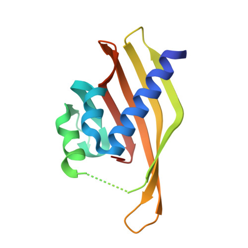
7URY - PubMed Abstract:
Proteins are long chains of amino acid residues that perform a myriad of functions in living organisms, including enzymatic reactions, signalling, and maintaining structural integrity. Protein function is determined directly by the protein structure. X-ray crystallography is the primary technique for determining the 3D structure of proteins, and facilitates understanding the effects of protein structure on function. The first step towards structure determination is crystallizing the protein of interest. We have developed a centrifugally-actuated microfluidic device that incorporates the fluid handling and metering necessary for protein crystallization. Liquid handling takes advantage of surface forces to control fluid flow and enable metering, without the need for any fluidic or pump connections. Our approach requires only the simple steps of pipetting the crystallization reagents into the device followed by either spinning or shaking to set up counter-diffusive protein crystallization trials. The use of thin, UV-curable polymers with a high level of X-ray transparency allows for in situ X-ray crystallography, eliminating the manual handling of fragile protein crystals and streamlining the process of protein structure analysis. We demonstrate the utility of our device using hen egg white lysozyme as a model system, followed by the crystallization and in situ , room temperature structural analysis of the hub domain of calcium-calmodulin dependent kinase II (CaMKIIβ).
- Department of Chemical Engineering, University of Massachusetts Amherst, MA 01003, USA. perrys@engin.umass.edu.
Organizational Affiliation: