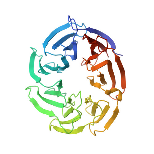
7SSE

Crystal structure of the WDR domain of human DCAF1 in complex with CYCA-117-70


Experimental Data Snapshot

  • Method: X-RAY DIFFRACTION
  • Resolution: 1.62 Å
  • R-Value Free: 0.234 
  • R-Value Work: 0.206 
  • R-Value Observed: 0.207 

wwPDB Validation   3D Report Full Report


Ligand Structure Quality Assessment 


This is version 1.3 of the entry. See complete history


Literature

Discovery of a Novel DCAF1 Ligand Using a Drug-Target Interaction Prediction Model: Generalizing Machine Learning to New Drug Targets.

Kimani, S.W.Owen, J.Green, S.R.Li, F.Li, Y.Dong, A.Brown, P.J.Ackloo, S.Kuter, D.Yang, C.MacAskill, M.MacKinnon, S.S.Arrowsmith, C.H.Schapira, M.Shahani, V.Halabelian, L.

(2023) J Chem Inf Model 63: 4070-4078

  • DOI: https://doi.org/10.1021/acs.jcim.3c00082
  • Primary Citation of Related Structures:  
    7SSE

  • PubMed Abstract: 

    DCAF1 functions as a substrate recruitment subunit for the RING-type CRL4 DCAF1 and the HECT family EDVP DCAF1 E3 ubiquitin ligases. The WDR domain of DCAF1 serves as a binding platform for substrate proteins and is also targeted by HIV and SIV lentiviral adaptors to induce the ubiquitination and proteasomal degradation of antiviral host factors. It is therefore attractive both as a potential therapeutic target for the development of chemical inhibitors and as an E3 ligase that could be recruited by novel PROTACs for targeted protein degradation. In this study, we used a proteome-scale drug-target interaction prediction model, MatchMaker, combined with cheminformatics filtering and docking to identify ligands for the DCAF1 WDR domain. Biophysical screening and X-ray crystallographic studies of the predicted binders confirmed a selective ligand occupying the central cavity of the WDR domain. This study shows that artificial intelligence-enabled virtual screening methods can successfully be applied in the absence of previously known ligands.


  • Organizational Affiliation

    Structural Genomics Consortium, University of Toronto, Toronto, Ontario M5G 1L7, Canada.


Macromolecules
Find similar proteins by:  (by identity cutoff)  |  3D Structure
Entity ID: 1
MoleculeChains Sequence LengthOrganismDetailsImage
DDB1- and CUL4-associated factor 1
A, B
315Homo sapiensMutation(s): 2 
Gene Names: DCAF1KIAA0800RIPVPRBP
EC: 2.7.11.1
UniProt & NIH Common Fund Data Resources
Find proteins for Q9Y4B6 (Homo sapiens)
Explore Q9Y4B6 
Go to UniProtKB:  Q9Y4B6
PHAROS:  Q9Y4B6
GTEx:  ENSG00000145041 
Entity Groups  
Sequence Clusters30% Identity50% Identity70% Identity90% Identity95% Identity100% Identity
UniProt GroupQ9Y4B6
Sequence Annotations
Expand
  • Reference Sequence
Small Molecules
Ligands 1 Unique
IDChains Name / Formula / InChI Key2D Diagram3D Interactions
B1I (Subject of Investigation/LOI)
Query on B1I

Download Ideal Coordinates CCD File 
C [auth A]N-[(3R)-1-(3-fluorophenyl)piperidin-3-yl]-6-(morpholin-4-yl)pyrimidin-4-amine
C19 H24 F N5 O
JEUVMKMFSZNWAD-MRXNPFEDSA-N
Experimental Data & Validation

Experimental Data

  • Method: X-RAY DIFFRACTION
  • Resolution: 1.62 Å
  • R-Value Free: 0.234 
  • R-Value Work: 0.206 
  • R-Value Observed: 0.207 
  • Space Group: P 1 21 1
Unit Cell:
Length ( Å )Angle ( ˚ )
a = 48.956α = 90
b = 87.919β = 98.72
c = 73.878γ = 90
Software Package:
Software NamePurpose
REFMACrefinement
SCALEPACKdata scaling
PDB_EXTRACTdata extraction
HKL-3000data reduction
PHASERphasing

Structure Validation

View Full Validation Report


Ligand Structure Quality Assessment 


Entry History & Funding Information

Deposition Data


Funding OrganizationLocationGrant Number
The Structural Genomics Consortium (SGC)Canada--

Revision History  (Full details and data files)

  • Version 1.0: 2021-12-15
    Type: Initial release
  • Version 1.1: 2022-02-23
    Changes: Database references, Source and taxonomy, Structure summary
  • Version 1.2: 2023-10-18
    Changes: Data collection, Refinement description
  • Version 1.3: 2023-11-22
    Changes: Database references