Crystal structure of plastocyanin from a green alga, Enteromorpha prolifera.
Collyer, C.A., Guss, J.M., Sugimura, Y., Yoshizaki, F., Freeman, H.C.(1990) J Mol Biol 211: 617-632
- PubMed: 2308169
- DOI: https://doi.org/10.1016/0022-2836(90)90269-R
- Primary Citation of Related Structures:
7PCY - PubMed Abstract:
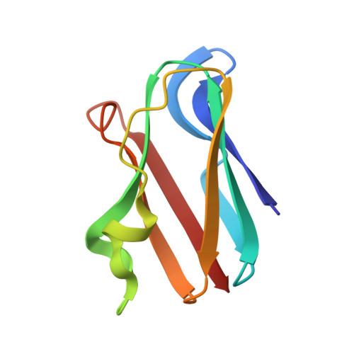
The crystal structure of the Cu-containing protein plastocyanin (Mr 10,500) from the green alga Enteromorpha prolifera has been solved by molecular replacement. The structure was refined by constrained-restrained and restrained reciprocal space least-squares techniques. The refined model includes 111 solvent sites. There is evidence for alternate conformers at eight residues. The residual is 0.12 for a data set comprising 74% of all observations accessible at 1.85 A resolution. The beta-sandwich structure of the algal plastocyanin is effectively the same as that of poplar leaf (Populus nigra var. italica) plastocyanin determined at 1.6 A resolution. The sequence homology between the two proteins is 56%. Differences between the contacts in the hydrophobic core create some significant (0.5 to 1.2 A) movements of the polypeptide backbone, resulting in small differences between the orientations and separations of corresponding beta-strands. These differences are most pronounced at the end of the molecule remote from the Cu site. The largest structural differences occur in the single non-beta strand, which includes the sole turn of helix in the molecule: two of the residues in a prominent kink of the poplar plastocyanin backbone are missing from the algal plastocyanin sequence, and there is a significant change in the position of the helical segment in relation to the beta-sandwich. Several other small but significant structural differences can be correlated with intermolecular contacts in the crystals. An intramolecular carboxyl-carboxylate hydrogen bond in the algal plastocyanin may be associated with an unusually high pKa. The dimensions of the Cu site in the two plastocyanins are, within the limits of precision, identical.
Organizational Affiliation:
Department of Inorganic Chemistry, University of Sydney, Australia.