7NW3

X-ray crystallographic study of PIYDIN, which contains the truncation determinants of binding PI and N, bound to RoAb13, a CCR5 antibody


Experimental Data Snapshot

  • Method: X-RAY DIFFRACTION
  • Resolution: 3.20 Å
  • R-Value Free: 0.293 
  • R-Value Work: 0.268 
  • R-Value Observed: 0.270 

wwPDB Validation   3D Report Full Report


This is version 1.2 of the entry. See complete history


Literature

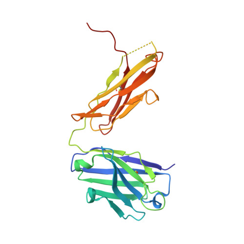
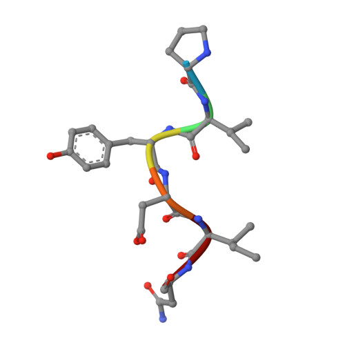
X-ray crystallographic studies of RoAb13 bound to PIYDIN, a part of the N-terminal domain of C-C chemokine receptor 5.

Govada, L.Saridakis, E.Kassen, S.C.Bin-Ramzi, A.Morgan, R.M.Chain, B.Helliwell, J.R.Chayen, N.E.

(2021) IUCrJ 8: 678-683

  • DOI: https://doi.org/10.1107/S2052252521005340
  • Primary Citation of Related Structures:  
    7NJZ, 7NW3

  • PubMed Abstract: 

    C-C chemokine receptor 5 (CCR5) is a major co-receptor molecule used by HIV-1 to enter cells. This led to the hypothesis that stimulating an antibody response would block HIV with minimal toxicity. Here, X-ray crystallographic studies of the anti-CCR5 antibody RoAb13 together with two peptides were undertaken: one peptide is a 31-residue peptide containing the PIYDIN sequence and the other is the PIDYIN peptide alone, where PIYDIN is part of the N-terminal region of CCR5 previously shown to be important for HIV entry. In the presence of the longer peptide (the complete N-terminal domain), difference electron density was observed at a site within a hypervariable CDR3 binding region. In the presence of the shorter core peptide PIYDIN, difference electron density is again observed at this CDR3 site, confirming consistent binding for both peptides. This may be useful in the design of a new biomimetic to stimulate an antibody response to CCR5 in order to block HIV infection.


  • Organizational Affiliation

    Division of Systems Medicine, Department of Metabolism, Digestion and Reproduction, Faculty of Medicine, Sir Alexander Fleming Building, Imperial College London, London SW7 2AZ, United Kingdom.


Macromolecules
Find similar proteins by:  (by identity cutoff)  |  3D Structure
Entity ID: 1
MoleculeChains Sequence LengthOrganismDetailsImage
Antibody RoAb13 Heavy ChainA [auth H]224Mus musculusMutation(s): 0 
Entity Groups  
Sequence Clusters30% Identity50% Identity70% Identity90% Identity95% Identity100% Identity
Sequence Annotations
Expand
  • Reference Sequence
Find similar proteins by:  (by identity cutoff)  |  3D Structure
Entity ID: 2
MoleculeChains Sequence LengthOrganismDetailsImage
Antibody RoAb13 Light ChainB [auth L]220Mus musculusMutation(s): 0 
Entity Groups  
Sequence Clusters30% Identity50% Identity70% Identity90% Identity95% Identity100% Identity
Sequence Annotations
Expand
  • Reference Sequence

Find similar proteins by:  Sequence   |   3D Structure  

Entity ID: 3
MoleculeChains Sequence LengthOrganismDetailsImage
Region from C-C chemokine receptor type 5 N-terminal domainC [auth A]6Homo sapiensMutation(s): 0 
UniProt & NIH Common Fund Data Resources
Find proteins for P51681 (Homo sapiens)
Explore P51681 
Go to UniProtKB:  P51681
PHAROS:  P51681
GTEx:  ENSG00000160791 
Entity Groups  
UniProt GroupP51681
Sequence Annotations
Expand
  • Reference Sequence
Experimental Data & Validation

Experimental Data

Unit Cell:
Length ( Å )Angle ( ˚ )
a = 76.607α = 90
b = 76.607β = 90
c = 270.06γ = 90
Software Package:
Software NamePurpose
PHENIXrefinement
XDSdata reduction
SCALAdata scaling
PHASERphasing

Structure Validation

View Full Validation Report


Entry History & Funding Information

Deposition Data


Funding OrganizationLocationGrant Number
Engineering and Physical Sciences Research CouncilUnited KingdomEP/K503733/1

Revision History  (Full details and data files)

  • Version 1.0: 2021-07-21
    Type: Initial release
  • Version 1.1: 2021-07-28
    Changes: Database references
  • Version 1.2: 2024-01-31
    Changes: Data collection, Database references, Refinement description